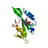
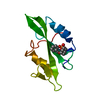
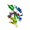
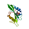
Journal: Mol Cell / Year: 2007 Title: Structural aspects of RbfA action during small ribosomal subunit assembly. Authors: Partha P Datta / Daniel N Wilson / Masahito Kawazoe / Neil K Swami / Tatsuya Kaminishi / Manjuli R Sharma / Timothy M Booth / Chie Takemoto / Paola Fucini / Shigeyuki Yokoyama / Rajendra K Agrawal / Abstract: Ribosome binding factor A (RbfA) is a bacterial cold shock response protein, required for an efficient processing of the 5' end of the 16S ribosomal RNA (rRNA) during assembly of the small (30S) ...Ribosome binding factor A (RbfA) is a bacterial cold shock response protein, required for an efficient processing of the 5' end of the 16S ribosomal RNA (rRNA) during assembly of the small (30S) ribosomal subunit. Here we present a crystal structure of Thermus thermophilus (Tth) RbfA and a three-dimensional cryo-electron microscopic (EM) map of the Tth 30S*RbfA complex. RbfA binds to the 30S subunit in a position overlapping the binding sites of the A and P site tRNAs, and RbfA's functionally important C terminus extends toward the 5' end of the 16S rRNA. In the presence of RbfA, a portion of the 16S rRNA encompassing helix 44, which is known to be directly involved in mRNA decoding and tRNA binding, is displaced. These results shed light on the role played by RbfA during maturation of the 30S subunit, and also indicate how RbfA provides cells with a translational advantage under conditions of cold shock.
History
Deposition
Sep 14, 2006
Deposition site: PDBJ / Processing site: PDBJ
Revision 1.0
Mar 14, 2007
Provider: repository / Type: Initial release
Revision 1.1
Mar 11, 2008
Group: Version format compliance
Revision 1.2
Jul 13, 2011
Group: Source and taxonomy / Version format compliance
In the structure databanks used in Yorodumi, some data are registered as the other names, "COVID-19 virus" and "2019-nCoV". Here are the details of the virus and the list of structure data.
Jan 31, 2019. EMDB accession codes are about to change! (news from PDBe EMDB page)
EMDB accession codes are about to change! (news from PDBe EMDB page)
The allocation of 4 digits for EMDB accession codes will soon come to an end. Whilst these codes will remain in use, new EMDB accession codes will include an additional digit and will expand incrementally as the available range of codes is exhausted. The current 4-digit format prefixed with “EMD-” (i.e. EMD-XXXX) will advance to a 5-digit format (i.e. EMD-XXXXX), and so on. It is currently estimated that the 4-digit codes will be depleted around Spring 2019, at which point the 5-digit format will come into force.
The EM Navigator/Yorodumi systems omit the EMD- prefix.
Related info.:Q: What is EMD? / ID/Accession-code notation in Yorodumi/EM Navigator
Yorodumi is a browser for structure data from EMDB, PDB, SASBDB, etc.
This page is also the successor to EM Navigator detail page, and also detail information page/front-end page for Omokage search.
The word "yorodu" (or yorozu) is an old Japanese word meaning "ten thousand". "mi" (miru) is to see.
Related info.:EMDB / PDB / SASBDB / Comparison of 3 databanks / Yorodumi Search / Aug 31, 2016. New EM Navigator & Yorodumi / Yorodumi Papers / Jmol/JSmol / Function and homology information / Changes in new EM Navigator and Yorodumi
 Movie
Movie Controller

 Yorodumi
 Open data
 Basic information
 Components
 Keywords
 RIBOSOMAL PROTEIN / 16S rRNA processing / 17S RNA /
  Function and homology information

 Authors
 Citation
 Structure visualization
 Downloads & links
 Other downloads
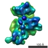


 PDBj
PDBj Assembly

 Mass: 18.015 Da / Num. of mol.: 105 / Source method: isolated from a natural source / Formula: H2O
 Sample preparation
 / Beamline: BL-5A / Wavelength: 1 Å
 Processing