 Movie
Movie Controller

[English] 日本語
 Yorodumi
- PDB-2dwk: Crystal structure of the RUN domain of mouse Rap2 interacting pro... -
+ Open data

- Basic information
| Entry | Database: PDB / ID: 2dwk | |||||||||
|---|---|---|---|---|---|---|---|---|---|---|
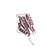
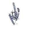
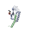
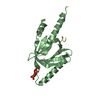
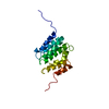
| Title | Crystal structure of the RUN domain of mouse Rap2 interacting protein x | |||||||||
 Components Components | Protein RUFY3 | |||||||||
 Keywords Keywords |  PROTEIN BINDING / RUN domain / effector / Rap2 / bundle / Structural Genomics / NPPSFA / National Project on Protein Structural and Functional Analyses / RIKEN Structural Genomics/Proteomics Initiative / RSGI PROTEIN BINDING / RUN domain / effector / Rap2 / bundle / Structural Genomics / NPPSFA / National Project on Protein Structural and Functional Analyses / RIKEN Structural Genomics/Proteomics Initiative / RSGI | |||||||||
| Function / homology |  Function and homology information Function and homology informationnegative regulation of axonogenesis / positive regulation of intracellular protein transport / anchoring junction / regulation of establishment of cell polarity / positive regulation of axonogenesis / regulation of axonogenesis / endomembrane system / positive regulation of axon extension / filopodium / actin filament organization ...negative regulation of axonogenesis / positive regulation of intracellular protein transport / anchoring junction / regulation of establishment of cell polarity / positive regulation of axonogenesis / regulation of axonogenesis / endomembrane system / positive regulation of axon extension / filopodium / actin filament organization / lamellipodium / nervous system development / growth cone / perikaryon / cell differentiation / positive regulation of cell migration / axon / neuronal cell body / dendrite / membrane / cytosol / cytoplasmSimilarity search - Function | |||||||||
| Biological species |  Mus musculus (house mouse) Mus musculus (house mouse) | |||||||||
| Method | X-RAY DIFFRACTION / SYNCHROTRON / MAD / Resolution: 2 Å | |||||||||
 Authors Authors | Kukimoto-Niino, M. / Murayama, K. / Shirouzu, M. / Yokoyama, S. / RIKEN Structural Genomics/Proteomics Initiative (RSGI) | |||||||||
 Citation Citation | Journal: J.Biol.Chem. / Year: 2006 Title: Crystal Structure of the RUN Domain of the RAP2-interacting Protein x Authors: Kukimoto-Niino, M. / Takagi, T. / Akasaka, R. / Murayama, K. / Uchikubo-Kamo, T. / Terada, T. / Inoue, M. / Watanabe, S. / Tanaka, A. / Hayashizaki, Y. / Kigawa, T. / Shirouzu, M. / Yokoyama, S. | |||||||||
| History |
|
- Structure visualization
| Structure viewer | Molecule: MolmilJmol/JSmol |
|---|
- Downloads & links
-Download
| PDBx/mmCIF format | 2dwk.cif.gz | 42.1 KB | Display | PDBx/mmCIF format |
|---|---|---|---|---|
| PDB format | pdb2dwk.ent.gz | 32.9 KB | Display | PDB format |
| PDBx/mmJSON format | 2dwk.json.gz | Tree view | PDBx/mmJSON format | |
| Others |  Other downloads Other downloads |
-Validation report
| Arichive directory | https://data.pdbj.org/pub/pdb/validation_reports/dw/2dwkftp://data.pdbj.org/pub/pdb/validation_reports/dw/2dwk | HTTPS FTP |
|---|
-Related structure data
| Related structure data |  2cxfC  2cxlC  2dwgC C: citing same article ( |
|---|---|
| Similar structure data | |
| Other databases |


-Links
 PDBj
PDBj- Assembly
| Deposited unit | 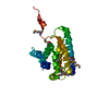
| ||||||||
|---|---|---|---|---|---|---|---|---|---|
| 1 |
| ||||||||
| Unit cell |
| ||||||||
| Components on special symmetry positions |
|
-Components
| #1: Protein | Mass: 20254.117 Da / Num. of mol.: 1 / Fragment: RUN domain Source method: isolated from a genetically manipulated source Source: (gene. exp.) Mus musculus (house mouse) / Description: Cell free protein synthesis / Gene: Rufy3, D5Bwg0860e, Ripx / Plasmid: PK011025-09 / Production host: Cell free synthesis / References: UniProt: Q9D394 |
|---|---|
| #2: Water | ChemComp-HOH / Water Mass: 18.015 Da / Num. of mol.: 68 / Source method: isolated from a natural source / Formula: H2O Mass: 18.015 Da / Num. of mol.: 68 / Source method: isolated from a natural source / Formula: H2O |
-Experimental details
-Experiment
| Experiment | Method: X-RAY DIFFRACTION / Number of used crystals: 1 |
|---|
- Sample preparation
| Crystal | Density Matthews: 2.84 Å3/Da / Density % sol: 56.73 % |
|---|---|
| Crystal grow | Temperature: 300 K / Method: vapor diffusion, hanging drop / pH: 8.5 Details: 0.05M SODIUM CHLORIDE, 1.4M AMMONIUM SULFATE, 0.1M TRIS, pH 8.5, VAPOR DIFFUSION, HANGING DROP, temperature 300K |
-Data collection
| Diffraction | Mean temperature: 100 K | ||||||||||||
|---|---|---|---|---|---|---|---|---|---|---|---|---|---|
| Diffraction source | Source: SYNCHROTRON / Site: SPring-8  / Beamline: BL26B1 / Wavelength: 0.9792, 0.9795, 0.9640 / Beamline: BL26B1 / Wavelength: 0.9792, 0.9795, 0.9640 | ||||||||||||
| Detector | Type: RIGAKU / Detector: IMAGE PLATE / Date: Nov 27, 2004 | ||||||||||||
| Radiation | Protocol: MAD / Monochromatic (M) / Laue (L): M / Scattering type: x-ray | ||||||||||||
| Radiation wavelength |
| ||||||||||||
| Reflection | Resolution: 2→50 Å / Num. obs: 16483 / % possible obs: 99.6 % / Observed criterion σ(I): -3 / Redundancy: 10.2 % / Biso Wilson estimate: 24.5 Å2 / Rsym value: 0.109 / Net I/σ(I): 14.5 | ||||||||||||
| Reflection shell | Resolution: 2→2.07 Å / Mean I/σ(I) obs: 5.2 / Rsym value: 0.497 / % possible all: 100 |
- Processing
| Software |
| ||||||||||||||||||||
|---|---|---|---|---|---|---|---|---|---|---|---|---|---|---|---|---|---|---|---|---|---|
| Refinement | Method to determine structure: MAD / Resolution: 2→33.59 Å / Rfactor Rfree error: 0.006 / Data cutoff high absF: 1897785.36 / Data cutoff low absF: 0 / Isotropic thermal model: RESTRAINED / Cross valid method: THROUGHOUT / σ(F): 0
| ||||||||||||||||||||
| Solvent computation | Solvent model: FLAT MODEL / Bsol: 55.5477 Å2 / ksol: 0.366412 e/Å3 | ||||||||||||||||||||
| Displacement parameters | Biso mean: 38.8 Å2
| ||||||||||||||||||||
| Refine analyze |
| ||||||||||||||||||||
| Refinement step | Cycle: LAST / Resolution: 2→33.59 Å
| ||||||||||||||||||||
| Refine LS restraints |
| ||||||||||||||||||||
| LS refinement shell | Resolution: 2→2.13 Å / Rfactor Rfree error: 0.018 / Total num. of bins used: 6
| ||||||||||||||||||||
| Xplor file |
|
