 Movie
Movie Controller

[English] 日本語
 Yorodumi
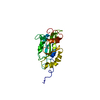
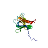
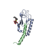
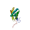
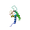
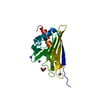
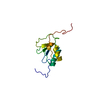
- PDB-1oqn: Crystal structure of the phosphotyrosine binding domain (PTB) of ... -
+ Open data

- Basic information
| Entry | Database: PDB / ID: 1oqn | ||||||
|---|---|---|---|---|---|---|---|
| Title | Crystal structure of the phosphotyrosine binding domain (PTB) of mouse Disabled 1 (Dab1) | ||||||
 Components Components |
| ||||||
 Keywords Keywords |  SIGNALING PROTEIN / PTB / Inositol / APP SIGNALING PROTEIN / PTB / Inositol / APP | ||||||
| Function / homology |  Function and homology information Function and homology informationcell-cell adhesion involved in neuronal-glial interactions involved in cerebral cortex radial glia guided migration / Formyl peptide receptors bind formyl peptides and many other ligands / lateral motor column neuron migration / TRAF6 mediated NF-kB activation / ECM proteoglycans / Advanced glycosylation endproduct receptor signaling / radial glia guided migration of Purkinje cell / amyloid-beta complex / Reelin signalling pathway / cerebral cortex radially oriented cell migration ...cell-cell adhesion involved in neuronal-glial interactions involved in cerebral cortex radial glia guided migration / Formyl peptide receptors bind formyl peptides and many other ligands / lateral motor column neuron migration / TRAF6 mediated NF-kB activation / ECM proteoglycans / Advanced glycosylation endproduct receptor signaling / radial glia guided migration of Purkinje cell / amyloid-beta complex / Reelin signalling pathway / cerebral cortex radially oriented cell migration / TAK1-dependent IKK and NF-kappa-B activation / growth cone lamellipodium / Post-translational protein phosphorylation / Regulation of Insulin-like Growth Factor (IGF) transport and uptake by Insulin-like Growth Factor Binding Proteins (IGFBPs) / cellular response to norepinephrine stimulus / Platelet degranulation / growth cone filopodium / endosome to plasma membrane transport vesicle / cerebellum structural organization / Golgi localization / positive regulation of endothelin production / ventral spinal cord development / radial glia-guided pyramidal neuron migration / Lysosome Vesicle Biogenesis / negative regulation of axonogenesis / Insertion of tail-anchored proteins into the endoplasmic reticulum membrane / motor neuron migration / lipoprotein particle / peptidase activator activity / growth factor receptor binding / regulation of amyloid-beta clearance / astrocyte projection / negative regulation of receptor signaling pathway via JAK-STAT / ion binding / regulation of epidermal growth factor-activated receptor activity / negative regulation of cell adhesion / frizzled binding / astrocyte differentiation / signaling receptor activator activity / collateral sprouting in absence of injury / cytosolic mRNA polyadenylation / microglia development / regulation of synapse structure or activity / axo-dendritic transport / synaptic assembly at neuromuscular junction / G alpha (q) signalling events / smooth endoplasmic reticulum calcium ion homeostasis / heparan sulfate proteoglycan binding / adult walking behavior / G alpha (i) signalling events / axon midline choice point recognition / astrocyte activation involved in immune response / regulation of spontaneous synaptic transmission / mating behavior / ciliary rootlet / main axon / PTB domain binding / Golgi-associated vesicle / low-density lipoprotein particle receptor binding / positive regulation of amyloid fibril formation / neuron remodeling / small GTPase-mediated signal transduction / : / nuclear envelope lumen / suckling behavior / presynaptic active zone / dendrite development / COPII-coated ER to Golgi transport vesicle / neuronal dense core vesicle / cerebral cortex cell migration / modulation of excitatory postsynaptic potential / cell surface receptor signaling pathway via JAK-STAT / apolipoprotein binding / brush border / neuromuscular process controlling balance / regulation of presynapse assembly / negative regulation of astrocyte differentiation / transition metal ion binding / intracellular copper ion homeostasis / regulation of multicellular organism growth / negative regulation of long-term synaptic potentiation / negative regulation of neuron differentiation / smooth endoplasmic reticulum / positive regulation of T cell migration / spindle midzone / cellular response to manganese ion / phosphatidylinositol 3-kinase binding / positive regulation of calcium-mediated signaling / forebrain development / clathrin-coated pit / cellular response to cAMP / regulation of peptidyl-tyrosine phosphorylation / positive regulation of chemokine production / Notch signaling pathway / cellular response to copper ion / positive regulation of G2/M transition of mitotic cell cycle / positive regulation of protein metabolic process / positive regulation of neuron differentiation / ionotropic glutamate receptor signaling pathway / neuron projection maintenanceSimilarity search - Function | ||||||
| Biological species |  Mus musculus (house mouse) Mus musculus (house mouse) | ||||||
| Method | X-RAY DIFFRACTION / SYNCHROTRON / MOLECULAR REPLACEMENT / Resolution: 2.3 Å | ||||||
 Authors Authors | Yun, M. / Keshvara, L. / Park, C.-G. / Zhang, Y.-M. / Dickerson, J.B. / Zheng, J. / Rock, C.O. / Curran, T. / Park, H.-W. | ||||||
 Citation Citation | Journal: J.Biol.Chem. / Year: 2003 Title: Crystal structures of the Dab homology domains of mouse disabled 1 and 2 Authors: Yun, M. / Keshvara, L. / Park, C.-G. / Zhang, Y.-M. / Dickerson, J.B. / Zheng, J. / Rock, C.O. / Curran, T. / Park, H.-W. | ||||||
| History |
|
- Structure visualization
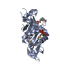
| Structure viewer | Molecule: MolmilJmol/JSmol |
|---|
- Downloads & links
-Download
| PDBx/mmCIF format | 1oqn.cif.gz | 80.8 KB | Display | PDBx/mmCIF format |
|---|---|---|---|---|
| PDB format | pdb1oqn.ent.gz | 61.1 KB | Display | PDB format |
| PDBx/mmJSON format | 1oqn.json.gz | Tree view | PDBx/mmJSON format | |
| Others |  Other downloads Other downloads |
-Validation report
| Arichive directory | https://data.pdbj.org/pub/pdb/validation_reports/oq/1oqnftp://data.pdbj.org/pub/pdb/validation_reports/oq/1oqn | HTTPS FTP |
|---|
-Related structure data
| Related structure data |  1m7eSC 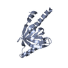 1p3rC S: Starting model for refinement C: citing same article ( |
|---|---|
| Similar structure data |

-Links
 PDBj
PDBj


- Assembly
| Deposited unit | 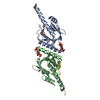
| ||||||||
|---|---|---|---|---|---|---|---|---|---|
| 1 | 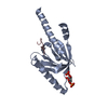
| ||||||||
| 2 | 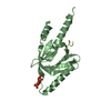
| ||||||||
| 3 |
| ||||||||
| Unit cell |
|
-Components
| #1: Protein | DAB1 / Dab1 Mass: 17960.758 Da / Num. of mol.: 2 / Fragment: PTB domain Source method: isolated from a genetically manipulated source Source: (gene. exp.) Mus musculus (house mouse) / Plasmid: pET30 / Species (production host): Escherichia coli / Production host:  Escherichia coli BL21(DE3) (bacteria) / Strain (production host): BL21(DE3) / References: UniProt: P97318 Escherichia coli BL21(DE3) (bacteria) / Strain (production host): BL21(DE3) / References: UniProt: P97318#2: Protein/peptide | Mass: 1086.132 Da / Num. of mol.: 2 / Fragment: 9mer peptide / Source method: obtained synthetically Details: The peptide was chemically synthesized. The sequence of the peptide is naturally found in Rattus norvegicus (rat). References: UniProt: P08592 #3: Chemical | Inositol trisphosphate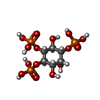  Mass: 420.096 Da / Num. of mol.: 2 / Source method: obtained synthetically / Formula: C6H15O15P3 Mass: 420.096 Da / Num. of mol.: 2 / Source method: obtained synthetically / Formula: C6H15O15P3#4: Water | ChemComp-HOH / | Water Mass: 18.015 Da / Num. of mol.: 217 / Source method: isolated from a natural source / Formula: H2O Mass: 18.015 Da / Num. of mol.: 217 / Source method: isolated from a natural source / Formula: H2O |
|---|
-Experimental details
-Experiment
| Experiment | Method: X-RAY DIFFRACTION / Number of used crystals: 1 |
|---|
- Sample preparation
| Crystal | Density Matthews: 2.33 Å3/Da / Density % sol: 47.31 % | ||||||||||||||||||||||||||||||||||||||||||
|---|---|---|---|---|---|---|---|---|---|---|---|---|---|---|---|---|---|---|---|---|---|---|---|---|---|---|---|---|---|---|---|---|---|---|---|---|---|---|---|---|---|---|---|
| Crystal grow | Temperature: 291 K / Method: vapor diffusion, hanging drop / pH: 8 Details: PEG 3350, magnesium sulfate, pH 8.0, VAPOR DIFFUSION, HANGING DROP, temperature 291K | ||||||||||||||||||||||||||||||||||||||||||
| Crystal grow | *PLUS Temperature: 18 ℃ / pH: 7.5 / Method: vapor diffusion, hanging drop | ||||||||||||||||||||||||||||||||||||||||||
| Components of the solutions | *PLUS
|
-Data collection
| Diffraction | Mean temperature: 100 K |
|---|---|
| Diffraction source | Source: SYNCHROTRON / Site: APS  / Beamline: 22-ID / Wavelength: 1.00465 Å / Beamline: 22-ID / Wavelength: 1.00465 Å |
| Detector | Type: MARRESEARCH / Detector: CCD / Date: Aug 20, 2002 |
| Radiation | Protocol: SINGLE WAVELENGTH / Monochromatic (M) / Laue (L): M / Scattering type: x-ray |
| Radiation wavelength | Wavelength: 1.00465 Å / Relative weight: 1 |
| Reflection | Resolution: 2.3→20 Å / Num. obs: 16118 / % possible obs: 99.5 % / Observed criterion σ(I): -3 / Redundancy: 24.8 % / Rsym value: 0.077 / Net I/σ(I): 27.3 |
| Reflection | *PLUS Num. measured all: 400319 / Rmerge(I) obs: 0.077 |
- Processing
| Software |
| ||||||||||||||||||||
|---|---|---|---|---|---|---|---|---|---|---|---|---|---|---|---|---|---|---|---|---|---|
| Refinement | Method to determine structure: MOLECULAR REPLACEMENT Starting model: PDB ENTRY 1M7E Resolution: 2.3→20 Å / Cross valid method: THROUGHOUT / σ(F): 2
| ||||||||||||||||||||
| Refinement step | Cycle: LAST / Resolution: 2.3→20 Å
| ||||||||||||||||||||
| Refine LS restraints |
| ||||||||||||||||||||
| Refinement | *PLUS Lowest resolution: 20 Å / % reflection Rfree: 5 % | ||||||||||||||||||||
| Solvent computation | *PLUS | ||||||||||||||||||||
| Displacement parameters | *PLUS | ||||||||||||||||||||
| Refine LS restraints | *PLUS
|
