 Movie
Movie Controller


+ Open data

- Basic information
| Entry | Database: PDB / ID: 2d02 | ||||||
|---|---|---|---|---|---|---|---|
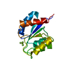
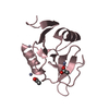
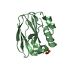
| Title | R52Q Mutant of Photoactive Yellow Protein, P65 Form | ||||||
 Components Components | Photoactive yellow protein | ||||||
 Keywords Keywords | SIGNALING PROTEIN / PAS / LOV / GAF-DOMAINS / PHOTORECEPTOR | ||||||
| Function / homology |  Function and homology informationphotoreceptor activity / phototransduction / regulation of DNA-templated transcription / identical protein binding Function and homology informationphotoreceptor activity / phototransduction / regulation of DNA-templated transcription / identical protein bindingSimilarity search - Function | ||||||
| Biological species |  Halorhodospira halophila (bacteria) Halorhodospira halophila (bacteria) | ||||||
| Method | X-RAY DIFFRACTION / SYNCHROTRON / MOLECULAR REPLACEMENT / Resolution: 1.42 Å | ||||||
 Authors Authors | Shimizu, N. / Kamikubo, H. / Yamazaki, Y. / Imamoto, Y. / Kataoka, M. | ||||||
 Citation Citation | Journal: Biochemistry / Year: 2006 Title: The Crystal Structure of the R52Q Mutant Demonstrates a Role for R52 in Chromophore pK(a) Regulation in Photoactive Yellow Protein Authors: Shimizu, N. / Kamikubo, H. / Yamazaki, Y. / Imamoto, Y. / Kataoka, M. | ||||||
| History |
|
- Structure visualization
| Structure viewer | Molecule: MolmilJmol/JSmol |
|---|
- Downloads & links
-Download
| PDBx/mmCIF format | 2d02.cif.gz | 72 KB | Display | PDBx/mmCIF format |
|---|---|---|---|---|
| PDB format | pdb2d02.ent.gz | 52.4 KB | Display | PDB format |
| PDBx/mmJSON format | 2d02.json.gz | Tree view | PDBx/mmJSON format | |
| Others |  Other downloads Other downloads |
-Validation report
| Arichive directory | https://data.pdbj.org/pub/pdb/validation_reports/d0/2d02ftp://data.pdbj.org/pub/pdb/validation_reports/d0/2d02 | HTTPS FTP |
|---|
-Related structure data
| Related structure data |  2d01C 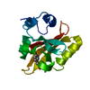 2phyS S: Starting model for refinement C: citing same article ( |
|---|---|
| Similar structure data |


-Links
 PDBj
PDBj


- Assembly
| Deposited unit | 
| ||||||||
|---|---|---|---|---|---|---|---|---|---|
| 1 |
| ||||||||
| Unit cell |
|
-Components
| #1: Protein | / Photoreceptor / PYP Mass: 13859.511 Da / Num. of mol.: 1 / Mutation: R52Q Source method: isolated from a genetically manipulated source Source: (gene. exp.) Halorhodospira halophila (bacteria) / Gene: PYP / Plasmid: PET / Species (production host): Escherichia coli / Production host: Escherichia coli BL21(DE3) (bacteria) / Strain (production host): BL21DE3 / References: UniProt: P16113 |
|---|---|
| #2: Chemical | ChemComp-HC4 / P-Coumaric acid  Mass: 164.158 Da / Num. of mol.: 1 / Source method: obtained synthetically / Formula: C9H8O3 Mass: 164.158 Da / Num. of mol.: 1 / Source method: obtained synthetically / Formula: C9H8O3 |
| #3: Water | ChemComp-HOH / Water Mass: 18.015 Da / Num. of mol.: 207 / Source method: isolated from a natural source / Formula: H2O Mass: 18.015 Da / Num. of mol.: 207 / Source method: isolated from a natural source / Formula: H2O |
-Experimental details
-Experiment
| Experiment | Method: X-RAY DIFFRACTION / Number of used crystals: 1 |
|---|
- Sample preparation
| Crystal | Density Matthews: 1.9 Å3/Da / Density % sol: 33.1 % |
|---|---|
| Crystal grow | Temperature: 293 K / Method: vapor diffusion / pH: 6 Details: 40% PEG2000, 100mM sodium citrate, pH 6.0, VAPOR DIFFUSION, temperature 293K |
-Data collection
| Diffraction | Mean temperature: 100 K |
|---|---|
| Diffraction source | Source: SYNCHROTRON / Site: SPring-8  / Beamline: BL40B2 / Wavelength: 1 Å / Beamline: BL40B2 / Wavelength: 1 Å |
| Detector | Type: ADSC QUANTUM 4 / Detector: CCD / Date: Jan 1, 2004 / Details: bending-magnet |
| Radiation | Monochromator: fixed-exit double crystal monochromator / Protocol: SINGLE WAVELENGTH / Monochromatic (M) / Laue (L): M / Scattering type: x-ray |
| Radiation wavelength | Wavelength: 1 Å / Relative weight: 1 |
| Reflection | Resolution: 1.42→25.74 Å / Num. obs: 18879 / % possible obs: 99.9 % / Redundancy: 11.1 % / Rmerge(I) obs: 0.07 / Net I/σ(I): 17 |
- Processing
| Software |
| |||||||||||||||||||||||||||||||||||||||||||||||||||||||||||||||||||||||||||||||||||||||||||||||||||||||||
|---|---|---|---|---|---|---|---|---|---|---|---|---|---|---|---|---|---|---|---|---|---|---|---|---|---|---|---|---|---|---|---|---|---|---|---|---|---|---|---|---|---|---|---|---|---|---|---|---|---|---|---|---|---|---|---|---|---|---|---|---|---|---|---|---|---|---|---|---|---|---|---|---|---|---|---|---|---|---|---|---|---|---|---|---|---|---|---|---|---|---|---|---|---|---|---|---|---|---|---|---|---|---|---|---|---|---|
| Refinement | Method to determine structure: MOLECULAR REPLACEMENT Starting model: 2PHY Resolution: 1.42→20 Å / Cor.coef. Fo:Fc: 0.971 / Cor.coef. Fo:Fc free: 0.97 / SU B: 1.906 / SU ML: 0.035 / Cross valid method: THROUGHOUT / ESU R: 0.085 / ESU R Free: 0.062 / Stereochemistry target values: MAXIMUM LIKELIHOOD / Details: HYDROGENS HAVE BEEN ADDED IN THE RIDING POSITIONS
| |||||||||||||||||||||||||||||||||||||||||||||||||||||||||||||||||||||||||||||||||||||||||||||||||||||||||
| Solvent computation | Ion probe radii: 0.8 Å / Shrinkage radii: 0.8 Å / VDW probe radii: 1.2 Å / Solvent model: MASK | |||||||||||||||||||||||||||||||||||||||||||||||||||||||||||||||||||||||||||||||||||||||||||||||||||||||||
| Displacement parameters | Biso mean: 13.247 Å2
| |||||||||||||||||||||||||||||||||||||||||||||||||||||||||||||||||||||||||||||||||||||||||||||||||||||||||
| Refinement step | Cycle: LAST / Resolution: 1.42→20 Å
| |||||||||||||||||||||||||||||||||||||||||||||||||||||||||||||||||||||||||||||||||||||||||||||||||||||||||
| Refine LS restraints |
| |||||||||||||||||||||||||||||||||||||||||||||||||||||||||||||||||||||||||||||||||||||||||||||||||||||||||
| LS refinement shell | Highest resolution: 1.42 Å / Num. reflection Rwork: 1285 / Total num. of bins used: 20 |
