 Movie
Movie Controller

[English] 日本語
 Yorodumi
- PDB-2cqq: Solution Structure of RSGI RUH-037, a myb DNA-binding domain in h... -
+ Open data

- Basic information
| Entry | Database: PDB / ID: 2cqq | ||||||
|---|---|---|---|---|---|---|---|
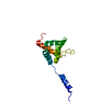
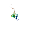
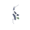
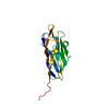
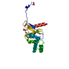
| Title | Solution Structure of RSGI RUH-037, a myb DNA-binding domain in human cDNA | ||||||
 Components Components | DnaJ homolog subfamily C member 1 | ||||||
 Keywords Keywords |  MEMBRANE PROTEIN / STRUCTURAL GENOMICS / NPPSFA / National Project on Protein Structural and Functional Analyses / RIKEN Structural Genomics/Proteomics Initiative / RSGI MEMBRANE PROTEIN / STRUCTURAL GENOMICS / NPPSFA / National Project on Protein Structural and Functional Analyses / RIKEN Structural Genomics/Proteomics Initiative / RSGI | ||||||
| Function / homology |  Function and homology informationregulation of protein secretion / ATPase activator activity / endomembrane system / negative regulation of proteolysis / protein folding / regulation of translation / protein-folding chaperone binding / nuclear membrane / endoplasmic reticulum membrane / endoplasmic reticulum ...regulation of protein secretion / ATPase activator activity / endomembrane system / negative regulation of proteolysis / protein folding / regulation of translation / protein-folding chaperone binding / nuclear membrane / endoplasmic reticulum membrane / endoplasmic reticulum / DNA binding / membrane / plasma membrane Function and homology informationregulation of protein secretion / ATPase activator activity / endomembrane system / negative regulation of proteolysis / protein folding / regulation of translation / protein-folding chaperone binding / nuclear membrane / endoplasmic reticulum membrane / endoplasmic reticulum ...regulation of protein secretion / ATPase activator activity / endomembrane system / negative regulation of proteolysis / protein folding / regulation of translation / protein-folding chaperone binding / nuclear membrane / endoplasmic reticulum membrane / endoplasmic reticulum / DNA binding / membrane / plasma membraneSimilarity search - Function | ||||||
| Biological species |  Homo sapiens (human) Homo sapiens (human) | ||||||
| Method | SOLUTION NMR / torsion angle dynamics | ||||||
 Authors Authors | Doi-Katayama, Y. / Hirota, H. / Hayashi, F. / Yokoyama, S. / RIKEN Structural Genomics/Proteomics Initiative (RSGI) | ||||||
 Citation Citation | Journal: To be Published Title: Solution Structure of RSGI RUH-037, a myb DNA-binding domain in human cDNA Authors: Doi-Katayama, Y. / Hirota, H. / Hayashi, F. / Yokoyama, S. | ||||||
| History |
|
- Structure visualization
| Structure viewer | Molecule: MolmilJmol/JSmol |
|---|
- Downloads & links
-Download
| PDBx/mmCIF format | 2cqq.cif.gz | 398.1 KB | Display | PDBx/mmCIF format |
|---|---|---|---|---|
| PDB format | pdb2cqq.ent.gz | 346.4 KB | Display | PDB format |
| PDBx/mmJSON format | 2cqq.json.gz | Tree view | PDBx/mmJSON format | |
| Others |  Other downloads Other downloads |
-Validation report
| Arichive directory | https://data.pdbj.org/pub/pdb/validation_reports/cq/2cqqftp://data.pdbj.org/pub/pdb/validation_reports/cq/2cqq | HTTPS FTP |
|---|
-Related structure data
| Similar structure data | |
|---|---|
| Other databases |

-Links
 PDBj
PDBj


- Assembly
| Deposited unit | 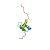
| |||||||||
|---|---|---|---|---|---|---|---|---|---|---|
| 1 |
| |||||||||
| NMR ensembles |
|
-Components
| #1: Protein | Mass: 7489.286 Da / Num. of mol.: 1 / Fragment: myb DNA-binding domain Source method: isolated from a genetically manipulated source Source: (gene. exp.) Homo sapiens (human) / Description: Cell-free protein synthesis / Gene: AY225122 / Plasmid: P041115-03 / References: UniProt: Q96KC8 |
|---|
-Experimental details
-Experiment
| Experiment | Method: SOLUTION NMR | ||||||||||||
|---|---|---|---|---|---|---|---|---|---|---|---|---|---|
| NMR experiment |
|
- Sample preparation
| Details | Contents: 1.55mM myb DNA-binding domain U-15N, 13C; 20mM d-Tris-HCl; 100mM NaCl; 1mM d-DTT; 0.02% NaN3; 90% H2O, 10% D2O Solvent system: 90% H2O/10% D2O |
|---|---|
| Sample conditions | Ionic strength: 120mM NaCl / pH: 7.0 / Pressure: ambient / Temperature: 298 K |
-NMR measurement
| NMR spectrometer | Type: Varian INOVA / Manufacturer: Varian / Model: INOVA / Field strength: 800 MHz |
|---|
- Processing
| NMR software |
| ||||||||||||||||||||||||||||
|---|---|---|---|---|---|---|---|---|---|---|---|---|---|---|---|---|---|---|---|---|---|---|---|---|---|---|---|---|---|
| Refinement | Method: torsion angle dynamics / Software ordinal: 1 | ||||||||||||||||||||||||||||
| NMR representative | Selection criteria: lowest energy | ||||||||||||||||||||||||||||
| NMR ensemble | Conformer selection criteria: target function, structures with the lowest energy, structures with the least restraint violations Conformers calculated total number: 100 / Conformers submitted total number: 20 |
