 Movie
Movie Controller


+ Open data

- Basic information
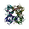
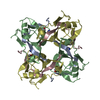
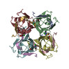
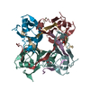
| Entry | Database: PDB / ID: 2c45 | ||||||
|---|---|---|---|---|---|---|---|
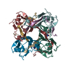
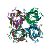
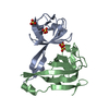
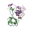
| Title | NATIVE PRECURSOR OF PYRUVOYL DEPENDENT ASPARTATE DECARBOXYLASE | ||||||
 Components Components | Aspartate 1-decarboxylase | ||||||
 Keywords Keywords | LYASE / DOUBLE-PSI BETA BARREL / CARBOXYLASE / ZYMOGEN / PANTOTHENATE BIOSYNTHESIS / DECARBOXYLASE / PYRUVATE | ||||||
| Function / homology |  Function and homology information Function and homology informationalanine biosynthetic process / aspartate 1-decarboxylase / aspartate 1-decarboxylase activity / pantothenate biosynthetic process / peptidoglycan-based cell wall / cytosolSimilarity search - Function | ||||||
| Biological species |   Mycobacterium tuberculosis (bacteria) Mycobacterium tuberculosis (bacteria) | ||||||
| Method | X-RAY DIFFRACTION / SYNCHROTRON / MOLECULAR REPLACEMENT / Resolution: 2.99 Å | ||||||
 Authors Authors | Gopalan, G. / Chopra, S. / Ranganathan, A. / Swaminathan, K. | ||||||
 Citation Citation | Journal: Proteins / Year: 2006 Title: Crystal structure of uncleaved L-aspartate-alpha-decarboxylase from Mycobacterium tuberculosis. Authors: Gopalan, G. / Chopra, S. / Ranganathan, A. / Swaminathan, K. #1: Journal: Nat.Struct.Biol. / Year: 1998Title: Crystal Structure of Aspartate Decarboxylase at 2.2A Resolution Provides Evidence for an Ester in Protein Self-Processing Authors: Albert, A. / Dhanaraj, V. / Genschel, U. / Khan, G. / Ramjee, M.K. / Pulido, R. / Sibanda, B.L. / von Delft, F. / Witty, M. / Blundell, T.L. / Smith, A.G. / Abell, C. #2: Journal: Embo J. / Year: 2003Title: Structural Constraints on Protein Self-Processing in L-Aspartate-Alpha-Decarboxylase Authors: Schmitzberger, F. / Kilkenny, M.L. / Lobley, C.M. / Webb, M.E. / Vinkovic, M. / Matak-Vinkovic, D. / Witty, M. / Chirgadze, D.Y. / Smith, A.G. / Abell, C. / Blundell, T.L. #3: Journal: J.Mol.Biol. / Year: 2004Title: Crystal Structure of the Schiff Base Intermediate Prior to Decarboxylation in the Catalytic Cycle of Aspartate Alpha-Decarboxylase Authors: Lee, B.I. / Suh, S.W. | ||||||
| History |
|
- Structure visualization
| Structure viewer | Molecule: MolmilJmol/JSmol |
|---|
- Downloads & links
-Download
| PDBx/mmCIF format | 2c45.cif.gz | 178.9 KB | Display | PDBx/mmCIF format |
|---|---|---|---|---|
| PDB format | pdb2c45.ent.gz | 142.4 KB | Display | PDB format |
| PDBx/mmJSON format | 2c45.json.gz | Tree view | PDBx/mmJSON format | |
| Others |  Other downloads Other downloads |
-Validation report
| Arichive directory | https://data.pdbj.org/pub/pdb/validation_reports/c4/2c45ftp://data.pdbj.org/pub/pdb/validation_reports/c4/2c45 | HTTPS FTP |
|---|
-Related structure data
| Related structure data |  1ppyS S: Starting model for refinement |
|---|---|
| Similar structure data |


-Links
 PDBj
PDBj


- Assembly
| Deposited unit | 
| ||||||||
|---|---|---|---|---|---|---|---|---|---|
| 1 | 
| ||||||||
| 2 | 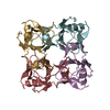
| ||||||||
| Unit cell |
|
-Components
| #1: Protein | / Aspartate alpha-decarboxylase Mass: 14900.986 Da / Num. of mol.: 8 Source method: isolated from a genetically manipulated source Source: (gene. exp.) Mycobacterium tuberculosis (bacteria) / Gene: panD, Rv3601c, MTCY07H7B.21 / Production host: Escherichia coli (E. coli) / References: UniProt: P9WIL3, aspartate 1-decarboxylase#2: Water | ChemComp-HOH / | Water Mass: 18.015 Da / Num. of mol.: 49 / Source method: isolated from a natural source / Formula: H2O Mass: 18.015 Da / Num. of mol.: 49 / Source method: isolated from a natural source / Formula: H2O |
|---|
-Experimental details
-Experiment
| Experiment | Method: X-RAY DIFFRACTION / Number of used crystals: 1 |
|---|
- Sample preparation
| Crystal | Density Matthews: 3.7 Å3/Da / Density % sol: 66.5 % |
|---|---|
| Crystal grow | Temperature: 292 K / Method: vapor diffusion, hanging drop / pH: 7.5 Details: SODIUM CACODYLATE (PH 6.5), 1.5 M MAGNESIUM SULPHATE AND 20% PEG2000, HANGING DROP, TEMPERATURE 292K |
-Data collection
| Diffraction | Mean temperature: 100 K |
|---|---|
| Diffraction source | Source: SYNCHROTRON / Site: NSLS  / Beamline: X12B / Wavelength: 1.1 / Beamline: X12B / Wavelength: 1.1 |
| Detector | Type: ADSC CCD / Detector: CCD / Date: Feb 14, 2004 |
| Radiation | Protocol: SINGLE WAVELENGTH / Monochromatic (M) / Laue (L): M / Scattering type: x-ray |
| Radiation wavelength | Wavelength: 1.1 Å / Relative weight: 1 |
| Reflection | Resolution: 2.99→50 Å / Num. obs: 30363 / % possible obs: 99.6 % / Observed criterion σ(I): 0 / Redundancy: 2.6 % / Biso Wilson estimate: 1.1 Å2 / Rmerge(I) obs: 0.12 |
| Reflection shell | Resolution: 2.99→3.1 Å / Redundancy: 2.6 % / % possible all: 96 |
- Processing
| Software |
| ||||||||||||||||||||||||||||||||||||||||||||||||||||||||||||
|---|---|---|---|---|---|---|---|---|---|---|---|---|---|---|---|---|---|---|---|---|---|---|---|---|---|---|---|---|---|---|---|---|---|---|---|---|---|---|---|---|---|---|---|---|---|---|---|---|---|---|---|---|---|---|---|---|---|---|---|---|---|
| Refinement | Method to determine structure: MOLECULAR REPLACEMENT Starting model: PDB ENTRY 1PPY Resolution: 2.99→7.99 Å / Rfactor Rfree error: 0.008 / Data cutoff high absF: 105724.32 / Isotropic thermal model: RESTRAINED / Cross valid method: THROUGHOUT / σ(F): 0 Details: THERE ARE EIGHT MOLECULES IN THE ASYMMETRIC UNIT, FORMING TWO TETRAMERS. HEMIHEDRAL TWINNING WITH TWIN FRACTION 0.437 AND TWIN OPERATOR H,-H-K,-L. RESTRAINED NCS MODE WAS USED FOR REFINEMENT.
| ||||||||||||||||||||||||||||||||||||||||||||||||||||||||||||
| Solvent computation | Solvent model: FLAT MODEL / Bsol: 49.9342 Å2 / ksol: 0.39434 e/Å3 | ||||||||||||||||||||||||||||||||||||||||||||||||||||||||||||
| Displacement parameters | Biso mean: 38.5 Å2
| ||||||||||||||||||||||||||||||||||||||||||||||||||||||||||||
| Refine analyze |
| ||||||||||||||||||||||||||||||||||||||||||||||||||||||||||||
| Refinement step | Cycle: LAST / Resolution: 2.99→7.99 Å
| ||||||||||||||||||||||||||||||||||||||||||||||||||||||||||||
| Refine LS restraints |
| ||||||||||||||||||||||||||||||||||||||||||||||||||||||||||||
| LS refinement shell | Resolution: 2.99→3.12 Å / Rfactor Rfree error: 0.035 / Total num. of bins used: 8
| ||||||||||||||||||||||||||||||||||||||||||||||||||||||||||||
| Xplor file |
|
