 Movie
Movie Controller

[English] 日本語
 Yorodumi
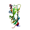
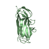
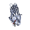
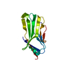
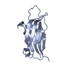
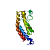
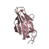
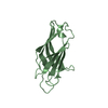
- PDB-2bwq: Crystal Structure of the RIM2 C2A-domain at 1.4 angstrom Resolution -
+ Open data

- Basic information
| Entry | Database: PDB / ID: 2bwq | ||||||
|---|---|---|---|---|---|---|---|
| Title | Crystal Structure of the RIM2 C2A-domain at 1.4 angstrom Resolution | ||||||
 Components Components | REGULATING SYNAPTIC MEMBRANE EXOCYTOSIS PROTEIN 2 | ||||||
 Keywords Keywords |  TRANSPORT PROTEIN / C2 DOMAIN / NEUROTRANSMITTER RELEASE TRANSPORT PROTEIN / C2 DOMAIN / NEUROTRANSMITTER RELEASE | ||||||
| Function / homology |  Function and homology information Function and homology informationregulation of calcium-dependent activation of synaptic vesicle fusion / maintenance of presynaptic active zone structure / structural constituent of presynaptic active zone / cytoskeleton of presynaptic active zone / positive regulation of inhibitory postsynaptic potential / spontaneous neurotransmitter secretion / synaptic vesicle docking / inhibitory synapse / calcium ion-regulated exocytosis of neurotransmitter / photoreceptor ribbon synapse ...regulation of calcium-dependent activation of synaptic vesicle fusion / maintenance of presynaptic active zone structure / structural constituent of presynaptic active zone / cytoskeleton of presynaptic active zone / positive regulation of inhibitory postsynaptic potential / spontaneous neurotransmitter secretion / synaptic vesicle docking / inhibitory synapse / calcium ion-regulated exocytosis of neurotransmitter / photoreceptor ribbon synapse / presynaptic active zone cytoplasmic component / calcium-ion regulated exocytosis / regulation of exocytosis / neurotransmitter secretion / positive regulation of dendrite extension / insulin secretion / synaptic vesicle priming / regulation of synaptic vesicle exocytosis / exocytosis / positive regulation of excitatory postsynaptic potential / GABA-ergic synapse / positive regulation of synaptic transmission / regulation of membrane potential / cell projection / establishment of localization in cell / intracellular protein transport / regulation of synaptic plasticity / adenylate cyclase-modulating G protein-coupled receptor signaling pathway / small GTPase binding / protein-macromolecule adaptor activity / presynaptic membrane / transmembrane transporter binding / cell differentiation / protein domain specific binding / glutamatergic synapse / protein-containing complex binding / positive regulation of gene expression / protein-containing complex / metal ion bindingSimilarity search - Function | ||||||
| Biological species |  RATTUS NORVEGICUS (Norway rat) RATTUS NORVEGICUS (Norway rat) | ||||||
| Method | X-RAY DIFFRACTION / SYNCHROTRON / MOLECULAR REPLACEMENT / Resolution: 1.41 Å | ||||||
 Authors Authors | Dai, H. / Tomchick, D.R. / Garcia, J. / Sudhof, T.C. / Machius, M. / Rizo, J. | ||||||
 Citation Citation | Journal: Biochemistry / Year: 2005 Title: Crystal Structure of the Rim2 C(2)A-Domain at 1.4 A Resolution. Authors: Dai, H. / Tomchick, D.R. / Garcia, J. / Sudhof, T.C. / Machius, M. / Rizo, J. | ||||||
| History |
|
- Structure visualization
| Structure viewer | Molecule: MolmilJmol/JSmol |
|---|
- Downloads & links
-Download
| PDBx/mmCIF format | 2bwq.cif.gz | 44.3 KB | Display | PDBx/mmCIF format |
|---|---|---|---|---|
| PDB format | pdb2bwq.ent.gz | 30.4 KB | Display | PDB format |
| PDBx/mmJSON format | 2bwq.json.gz | Tree view | PDBx/mmJSON format | |
| Others |  Other downloads Other downloads |
-Validation report
| Arichive directory | https://data.pdbj.org/pub/pdb/validation_reports/bw/2bwqftp://data.pdbj.org/pub/pdb/validation_reports/bw/2bwq | HTTPS FTP |
|---|
-Related structure data
| Related structure data |  1v27S S: Starting model for refinement |
|---|---|
| Similar structure data |


-Links
 PDBj
PDBj


- Assembly
| Deposited unit | 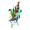
| ||||||||
|---|---|---|---|---|---|---|---|---|---|
| 1 |
| ||||||||
| Unit cell |
|
-Components
| #1: Protein | Mass: 15580.859 Da / Num. of mol.: 1 / Fragment: C2 DOMAIN, RESIDUES 725-853 Source method: isolated from a genetically manipulated source Source: (gene. exp.) RATTUS NORVEGICUS (Norway rat) / Plasmid: PGEX-KG / Production host:  ESCHERICHIA COLI (E. coli) / Strain (production host): BL21 / References: UniProt: Q9JIS1 ESCHERICHIA COLI (E. coli) / Strain (production host): BL21 / References: UniProt: Q9JIS1 | ||
|---|---|---|---|
| #2: Chemical | Sulfate  Mass: 96.063 Da / Num. of mol.: 2 / Source method: obtained synthetically / Formula: SO4 Mass: 96.063 Da / Num. of mol.: 2 / Source method: obtained synthetically / Formula: SO4#3: Water | ChemComp-HOH / | Water Mass: 18.015 Da / Num. of mol.: 110 / Source method: isolated from a natural source / Formula: H2O Mass: 18.015 Da / Num. of mol.: 110 / Source method: isolated from a natural source / Formula: H2O |
-Experimental details
-Experiment
| Experiment | Method: X-RAY DIFFRACTION / Number of used crystals: 1 |
|---|
- Sample preparation
| Crystal | Density Matthews: 1.8 Å3/Da / Density % sol: 29.7 % |
|---|---|
| Crystal grow | Temperature: 293 K / Method: vapor diffusion, hanging drop / pH: 4.5 Details: RAT RIM2 C2A-DOMAIN (RESIDUES 722-859) DISSOLVED IN 20 MM MES (PH 6.0), 150 MM NACL AND 1 MM EDTA WAS CONCENTRATED TO 25 MG/ML AND CRYSTALLIZED IN 17.5% (W/V) PEG 4000, 0.2 M (NH4)2SO4, 0.1M ...Details: RAT RIM2 C2A-DOMAIN (RESIDUES 722-859) DISSOLVED IN 20 MM MES (PH 6.0), 150 MM NACL AND 1 MM EDTA WAS CONCENTRATED TO 25 MG/ML AND CRYSTALLIZED IN 17.5% (W/V) PEG 4000, 0.2 M (NH4)2SO4, 0.1M SODIUM ACETATE (PH 4.5) AT 20 DEGREES C USING THE HANGING-DROP VAPOR-DIFFUSION METHOD. CRYSTALS APPEARED OVERNIGHT AND GREW TO A FINAL SIZE OF 0.05 X 0.05 X 0.1 MM WITHIN TWO DAYS. PRIOR TO DATA COLLECTION, CRYSTALS WERE TRANSFERRED INTO A SOLUTION OF 20% (W/V) PEG 4000, 0.15M NACL, 0.2 M (NH4)2SO4, 0.1M SODIUM ACETATE (PH 4.5) AND 15% (V/V) ETHYLENE GLYCOL, AND THEN FLASH-COOLED IN LIQUID PROPANE. |
-Data collection
| Diffraction | Mean temperature: 100 K |
|---|---|
| Diffraction source | Source: SYNCHROTRON / Site: APS  / Beamline: 19-ID / Wavelength: 1.00691 / Beamline: 19-ID / Wavelength: 1.00691 |
| Detector | Type: CUSTOM / Detector: CCD / Date: Aug 13, 2004 |
| Radiation | Protocol: SINGLE WAVELENGTH / Monochromatic (M) / Laue (L): M / Scattering type: x-ray |
| Radiation wavelength | Wavelength: 1.00691 Å / Relative weight: 1 |
| Reflection | Resolution: 1.41→20.91 Å / Num. obs: 23283 / % possible obs: 98.5 % / Observed criterion σ(I): -3 / Redundancy: 4 % / Rmerge(I) obs: 0.02 / Net I/σ(I): 52.9 |
| Reflection shell | Resolution: 1.41→1.43 Å / Redundancy: 2.8 % / Rmerge(I) obs: 0.18 / Mean I/σ(I) obs: 5.2 / % possible all: 82 |
- Processing
| Software |
| ||||||||||||||||||||||||||||||||||||||||||||||||||||||||||||||||||||||||||||||||||||||||||||||||||||||||||||||||||||||||||||||||||||||||||||||||||||||||||||||||||||||||||||||||||||||
|---|---|---|---|---|---|---|---|---|---|---|---|---|---|---|---|---|---|---|---|---|---|---|---|---|---|---|---|---|---|---|---|---|---|---|---|---|---|---|---|---|---|---|---|---|---|---|---|---|---|---|---|---|---|---|---|---|---|---|---|---|---|---|---|---|---|---|---|---|---|---|---|---|---|---|---|---|---|---|---|---|---|---|---|---|---|---|---|---|---|---|---|---|---|---|---|---|---|---|---|---|---|---|---|---|---|---|---|---|---|---|---|---|---|---|---|---|---|---|---|---|---|---|---|---|---|---|---|---|---|---|---|---|---|---|---|---|---|---|---|---|---|---|---|---|---|---|---|---|---|---|---|---|---|---|---|---|---|---|---|---|---|---|---|---|---|---|---|---|---|---|---|---|---|---|---|---|---|---|---|---|---|---|---|
| Refinement | Method to determine structure: MOLECULAR REPLACEMENT Starting model: PDB ENTRY 1V27 Resolution: 1.41→20 Å / Cor.coef. Fo:Fc: 0.961 / Cor.coef. Fo:Fc free: 0.949 / SU B: 2.046 / SU ML: 0.041 / TLS residual ADP flag: LIKELY RESIDUAL / Cross valid method: THROUGHOUT / ESU R: 0.071 / ESU R Free: 0.073 / Stereochemistry target values: MAXIMUM LIKELIHOOD / Details: HYDROGENS HAVE BEEN ADDED IN THE RIDING POSITIONS.
| ||||||||||||||||||||||||||||||||||||||||||||||||||||||||||||||||||||||||||||||||||||||||||||||||||||||||||||||||||||||||||||||||||||||||||||||||||||||||||||||||||||||||||||||||||||||
| Solvent computation | Ion probe radii: 0.8 Å / Shrinkage radii: 0.8 Å / VDW probe radii: 1.2 Å / Solvent model: MASK | ||||||||||||||||||||||||||||||||||||||||||||||||||||||||||||||||||||||||||||||||||||||||||||||||||||||||||||||||||||||||||||||||||||||||||||||||||||||||||||||||||||||||||||||||||||||
| Displacement parameters | Biso mean: 17.71 Å2
| ||||||||||||||||||||||||||||||||||||||||||||||||||||||||||||||||||||||||||||||||||||||||||||||||||||||||||||||||||||||||||||||||||||||||||||||||||||||||||||||||||||||||||||||||||||||
| Refinement step | Cycle: LAST / Resolution: 1.41→20 Å
| ||||||||||||||||||||||||||||||||||||||||||||||||||||||||||||||||||||||||||||||||||||||||||||||||||||||||||||||||||||||||||||||||||||||||||||||||||||||||||||||||||||||||||||||||||||||
| Refine LS restraints |
|