 Movie
Movie Controller

[English] 日本語
 Yorodumi
- PDB-2bu8: crystal structures of human pyruvate dehydrogenase kinase 2 conta... -
+ Open data

- Basic information
| Entry | Database: PDB / ID: 2bu8 | ||||||
|---|---|---|---|---|---|---|---|
| Title | crystal structures of human pyruvate dehydrogenase kinase 2 containing physiological and synthetic ligands | ||||||
 Components Components | PYRUVATE DEHYDROGENASE KINASE ISOENZYME 2 | ||||||
 Keywords Keywords |  TRANSFERASE / PYRUVATE DEHYDROGENASE KINASE 2 GHKL MOTIF REGULATION TRANSFERASE / PYRUVATE DEHYDROGENASE KINASE 2 GHKL MOTIF REGULATION | ||||||
| Function / homology |  Function and homology information Function and homology information[pyruvate dehydrogenase (acetyl-transferring)] kinase / regulation of acetyl-CoA biosynthetic process from pyruvate / pyruvate dehydrogenase (acetyl-transferring) kinase activity / : / regulation of cellular ketone metabolic process / regulation of pH / cellular response to nutrient / Regulation of pyruvate dehydrogenase (PDH) complex / Signaling by Retinoic Acid / regulation of gluconeogenesis ...[pyruvate dehydrogenase (acetyl-transferring)] kinase / regulation of acetyl-CoA biosynthetic process from pyruvate / pyruvate dehydrogenase (acetyl-transferring) kinase activity / : / regulation of cellular ketone metabolic process / regulation of pH / cellular response to nutrient / Regulation of pyruvate dehydrogenase (PDH) complex / Signaling by Retinoic Acid / regulation of gluconeogenesis / intrinsic apoptotic signaling pathway by p53 class mediator / regulation of glucose metabolic process / regulation of calcium-mediated signaling / cellular response to reactive oxygen species / glucose metabolic process / insulin receptor signaling pathway / glucose homeostasis / protein kinase activity / mitochondrial matrix / phosphorylation / protein homodimerization activity / mitochondrion / nucleoplasm / ATP binding / cytosolSimilarity search - Function | ||||||
| Biological species |  HOMO SAPIENS (human) HOMO SAPIENS (human) | ||||||
| Method | X-RAY DIFFRACTION / SYNCHROTRON / MOLECULAR REPLACEMENT / Resolution: 2.5 Å | ||||||
 Authors Authors | Knoechel, T.R. / Tucker, A.D. / Robinson, C.M. / Phillips, C. / Taylor, W. / Bungay, P.J. / Kasten, S.A. / Roche, T.E. / Brown, D.G. | ||||||
 Citation Citation | Journal: Biochemistry / Year: 2006 Title: Regulatory Roles of the N-Terminal Domain Based on Crystal Structures of Human Pyruvate Dehydrogenase Kinase 2 Containing Physiological and Synthetic Ligands. Authors: Knoechel, T.R. / Tucker, A.D. / Robinson, C.M. / Phillips, C. / Taylor, W. / Bungay, P.J. / Kasten, S.A. / Roche, T.E. / Brown, D.G. | ||||||
| History |
|
- Structure visualization
| Structure viewer | Molecule: MolmilJmol/JSmol |
|---|
- Downloads & links
-Download
| PDBx/mmCIF format | 2bu8.cif.gz | 91.3 KB | Display | PDBx/mmCIF format |
|---|---|---|---|---|
| PDB format | pdb2bu8.ent.gz | 67.5 KB | Display | PDB format |
| PDBx/mmJSON format | 2bu8.json.gz | Tree view | PDBx/mmJSON format | |
| Others |  Other downloads Other downloads |
-Validation report
| Arichive directory | https://data.pdbj.org/pub/pdb/validation_reports/bu/2bu8ftp://data.pdbj.org/pub/pdb/validation_reports/bu/2bu8 | HTTPS FTP |
|---|
-Related structure data
| Related structure data |  2btzSC  2bu2C  2bu5C  2bu6C  2bu7C S: Starting model for refinement C: citing same article ( |
|---|---|
| Similar structure data |


-Links
 PDBj
PDBj


- Assembly
| Deposited unit | 
| ||||||||
|---|---|---|---|---|---|---|---|---|---|
| 1 | 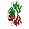
| ||||||||
| Unit cell |
|
-Components
| #1: Protein | Mass: 44647.730 Da / Num. of mol.: 1 Source method: isolated from a genetically manipulated source Source: (gene. exp.) HOMO SAPIENS (human) / Plasmid: PFASTBAC1 / Cell line (production host): High Five / Production host:  TRICHOPLUSIA NI (cabbage looper) / References: UniProt: Q15119, EC: 2.7.1.99 TRICHOPLUSIA NI (cabbage looper) / References: UniProt: Q15119, EC: 2.7.1.99 |
|---|---|
| #2: Chemical | ChemComp-ADP / Adenosine diphosphate  Mass: 427.201 Da / Num. of mol.: 1 / Source method: obtained synthetically / Formula: C10H15N5O10P2 / Comment: ADP, energy-carrying molecule*YM Mass: 427.201 Da / Num. of mol.: 1 / Source method: obtained synthetically / Formula: C10H15N5O10P2 / Comment: ADP, energy-carrying molecule*YM |
| #3: Chemical | ChemComp-MG /   Mass: 24.305 Da / Num. of mol.: 1 / Source method: obtained synthetically / Formula: Mg Mass: 24.305 Da / Num. of mol.: 1 / Source method: obtained synthetically / Formula: Mg |
| #4: Chemical | ChemComp-TF4 / Dichloroacetic acid  Mass: 128.942 Da / Num. of mol.: 1 / Source method: obtained synthetically / Formula: C2H2Cl2O2 Mass: 128.942 Da / Num. of mol.: 1 / Source method: obtained synthetically / Formula: C2H2Cl2O2 |
| #5: Water | ChemComp-HOH / Water Mass: 18.015 Da / Num. of mol.: 151 / Source method: isolated from a natural source / Formula: H2O Mass: 18.015 Da / Num. of mol.: 151 / Source method: isolated from a natural source / Formula: H2O |
| Sequence details | N -TERMINAL GS CLONING ARTEFACT |
-Experimental details
-Experiment
| Experiment | Method: X-RAY DIFFRACTION / Number of used crystals: 1 |
|---|
- Sample preparation
| Crystal | Density Matthews: 3.35 Å3/Da / Density % sol: 62 % |
|---|---|
| Crystal grow | Temperature: 277 K / Method: vapor diffusion, hanging drop / pH: 6 Details: 100MM MES PH5.8-6,10% ISOPROPANOL,200MM CALCIUM ACETATE,10MG/ML PROTEIN,4 DEGREES, pH 6.00 |
-Data collection
| Diffraction | Mean temperature: 100 K |
|---|---|
| Diffraction source | Source: SYNCHROTRON / Site: SRS  / Beamline: PX9.6 / Wavelength: 0.87 / Beamline: PX9.6 / Wavelength: 0.87 |
| Detector | Type: ADSC CCD / Detector: CCD / Details: MIRRORS |
| Radiation | Protocol: SINGLE WAVELENGTH / Monochromatic (M) / Laue (L): M / Scattering type: x-ray |
| Radiation wavelength | Wavelength: 0.87 Å / Relative weight: 1 |
| Reflection | Resolution: 2.5→30 Å / Num. obs: 21614 / % possible obs: 99.2 % / Observed criterion σ(I): 0 / Redundancy: 3.5 % / Rmerge(I) obs: 0.04 |
| Reflection shell | Resolution: 2.5→2.59 Å / Redundancy: 3.1 % / Rmerge(I) obs: 0.23 / % possible all: 98.9 |
- Processing
| Software |
| ||||||||||||||||||||||||||||||||||||||||||||||||||||||||||||
|---|---|---|---|---|---|---|---|---|---|---|---|---|---|---|---|---|---|---|---|---|---|---|---|---|---|---|---|---|---|---|---|---|---|---|---|---|---|---|---|---|---|---|---|---|---|---|---|---|---|---|---|---|---|---|---|---|---|---|---|---|---|
| Refinement | Method to determine structure: MOLECULAR REPLACEMENT Starting model: PDB ENTRY 2BTZ Resolution: 2.5→30 Å / Cross valid method: THROUGHOUT / σ(F): 0
| ||||||||||||||||||||||||||||||||||||||||||||||||||||||||||||
| Solvent computation | Solvent model: BABINET / Bsol: 73.5571 Å2 / ksol: 0.355715 e/Å3 | ||||||||||||||||||||||||||||||||||||||||||||||||||||||||||||
| Displacement parameters |
| ||||||||||||||||||||||||||||||||||||||||||||||||||||||||||||
| Refinement step | Cycle: LAST / Resolution: 2.5→30 Å
| ||||||||||||||||||||||||||||||||||||||||||||||||||||||||||||
| Refine LS restraints |
| ||||||||||||||||||||||||||||||||||||||||||||||||||||||||||||
| Xplor file |
|
