 Movie
Movie Controller


+ Open data

- Basic information
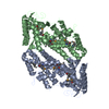
| Entry | Database: PDB / ID: 4mdv | ||||||
|---|---|---|---|---|---|---|---|
| Title | Crystal structure of calcium-bound annexin (Sm)1 | ||||||
 Components Components | Annexin | ||||||
 Keywords Keywords | METAL BINDING PROTEIN / annexin / calcium-binding protein | ||||||
| Function / homology |  Function and homology information Function and homology informationcalcium-dependent phospholipid binding / calcium ion binding / identical protein bindingSimilarity search - Function | ||||||
| Biological species |  Schistosoma mansoni (invertebrata) Schistosoma mansoni (invertebrata) | ||||||
| Method | X-RAY DIFFRACTION / SYNCHROTRON / MOLECULAR REPLACEMENT / Resolution: 2.5 Å | ||||||
 Authors Authors | Hofmann, A. | ||||||
 Citation Citation | Journal: Febs J. / Year: 2014 Title: Crystal structure and immunological properties of the first annexin from Schistosoma mansoni: insights into the structural integrity of the schistosomal tegument. Authors: Leow, C.Y. / Willis, C. / Osman, A. / Mason, L. / Simon, A. / Smith, B.J. / Gasser, R.B. / Jones, M.K. / Hofmann, A. | ||||||
| History |
|
- Structure visualization
| Structure viewer | Molecule: MolmilJmol/JSmol |
|---|
- Downloads & links
-Download
| PDBx/mmCIF format | 4mdv.cif.gz | 301.6 KB | Display | PDBx/mmCIF format |
|---|---|---|---|---|
| PDB format | pdb4mdv.ent.gz | 243.2 KB | Display | PDB format |
| PDBx/mmJSON format | 4mdv.json.gz | Tree view | PDBx/mmJSON format | |
| Others |  Other downloads Other downloads |
-Validation report
| Arichive directory | https://data.pdbj.org/pub/pdb/validation_reports/md/4mdvftp://data.pdbj.org/pub/pdb/validation_reports/md/4mdv | HTTPS FTP |
|---|
-Related structure data
| Related structure data | 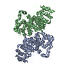 4mduSC S: Starting model for refinement C: citing same article ( |
|---|---|
| Similar structure data |


-Links
 PDBj
PDBj- Assembly
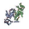
| Deposited unit | 
| ||||||||||||||||||
|---|---|---|---|---|---|---|---|---|---|---|---|---|---|---|---|---|---|---|---|
| 1 |
| ||||||||||||||||||
| 2 |
| ||||||||||||||||||
| Unit cell |
| ||||||||||||||||||
| Noncrystallographic symmetry (NCS) | NCS domain:
NCS domain segments:
|
-Components
| #1: Protein | Mass: 42815.691 Da / Num. of mol.: 2 Source method: isolated from a genetically manipulated source Source: (gene. exp.) Schistosoma mansoni (invertebrata) / Gene: annexin (Sm)1, Smp_074150.1 / Plasmid: pET28 / Production host:  Escherichia coli (E. coli) / Strain (production host): BL21(DE3) / References: UniProt: C4QH88 Escherichia coli (E. coli) / Strain (production host): BL21(DE3) / References: UniProt: C4QH88#2: Chemical | ChemComp-CA /   Mass: 40.078 Da / Num. of mol.: 13 / Source method: obtained synthetically / Formula: Ca Mass: 40.078 Da / Num. of mol.: 13 / Source method: obtained synthetically / Formula: Ca#3: Water | ChemComp-HOH / | Water Mass: 18.015 Da / Num. of mol.: 117 / Source method: isolated from a natural source / Formula: H2O Mass: 18.015 Da / Num. of mol.: 117 / Source method: isolated from a natural source / Formula: H2O |
|---|
-Experimental details
-Experiment
| Experiment | Method: X-RAY DIFFRACTION / Number of used crystals: 1 |
|---|
- Sample preparation
| Crystal | Density Matthews: 2.25 Å3/Da / Density % sol: 45.28 % |
|---|---|
| Crystal grow | Temperature: 289 K / Method: vapor diffusion, hanging drop / pH: 9 Details: 12.5% PEG2000, 50 mM TRIS, VAPOR DIFFUSION, HANGING DROP, temperature 289K, pH 9.0 |
-Data collection
| Diffraction | Mean temperature: 100 K |
|---|---|
| Diffraction source | Source: SYNCHROTRON / Site: Australian Synchrotron  / Beamline: MX1 / Wavelength: 1.3378 Å / Beamline: MX1 / Wavelength: 1.3378 Å |
| Detector | Type: ADSC QUANTUM 210r / Detector: CCD / Date: Aug 8, 2013 |
| Radiation | Protocol: SINGLE WAVELENGTH / Monochromatic (M) / Laue (L): M / Scattering type: x-ray |
| Radiation wavelength | Wavelength: 1.3378 Å / Relative weight: 1 |
| Reflection | Resolution: 2.5→24.1 Å / Num. obs: 26180 / % possible obs: 99.8 % / Redundancy: 3.8 % / Rsym value: 0.041 |
| Reflection shell | Highest resolution: 2.5 Å / Redundancy: 3.6 % / Rsym value: 0.386 / % possible all: 99.5 |
- Processing
| Software |
| ||||||||||||||||||||||||||||||||||||||||||||||||||||||||||||||||||||||||||||||||||||||||||||||||||||||||||||||||||||||||||||||||||||||||||||
|---|---|---|---|---|---|---|---|---|---|---|---|---|---|---|---|---|---|---|---|---|---|---|---|---|---|---|---|---|---|---|---|---|---|---|---|---|---|---|---|---|---|---|---|---|---|---|---|---|---|---|---|---|---|---|---|---|---|---|---|---|---|---|---|---|---|---|---|---|---|---|---|---|---|---|---|---|---|---|---|---|---|---|---|---|---|---|---|---|---|---|---|---|---|---|---|---|---|---|---|---|---|---|---|---|---|---|---|---|---|---|---|---|---|---|---|---|---|---|---|---|---|---|---|---|---|---|---|---|---|---|---|---|---|---|---|---|---|---|---|---|---|
| Refinement | Method to determine structure: MOLECULAR REPLACEMENT Starting model: 4MDU Resolution: 2.5→24.1 Å / SU ML: 0.47 / σ(F): 0.98 / Phase error: 28.21 / Stereochemistry target values: ML
| ||||||||||||||||||||||||||||||||||||||||||||||||||||||||||||||||||||||||||||||||||||||||||||||||||||||||||||||||||||||||||||||||||||||||||||
| Solvent computation | Shrinkage radii: 0.9 Å / VDW probe radii: 1.11 Å / Solvent model: FLAT BULK SOLVENT MODEL / Bsol: 70.315 Å2 / ksol: 0.349 e/Å3 | ||||||||||||||||||||||||||||||||||||||||||||||||||||||||||||||||||||||||||||||||||||||||||||||||||||||||||||||||||||||||||||||||||||||||||||
| Displacement parameters |
| ||||||||||||||||||||||||||||||||||||||||||||||||||||||||||||||||||||||||||||||||||||||||||||||||||||||||||||||||||||||||||||||||||||||||||||
| Refinement step | Cycle: LAST / Resolution: 2.5→24.1 Å
| ||||||||||||||||||||||||||||||||||||||||||||||||||||||||||||||||||||||||||||||||||||||||||||||||||||||||||||||||||||||||||||||||||||||||||||
| Refine LS restraints |
| ||||||||||||||||||||||||||||||||||||||||||||||||||||||||||||||||||||||||||||||||||||||||||||||||||||||||||||||||||||||||||||||||||||||||||||
| Refine LS restraints NCS |
| ||||||||||||||||||||||||||||||||||||||||||||||||||||||||||||||||||||||||||||||||||||||||||||||||||||||||||||||||||||||||||||||||||||||||||||
| LS refinement shell |
| ||||||||||||||||||||||||||||||||||||||||||||||||||||||||||||||||||||||||||||||||||||||||||||||||||||||||||||||||||||||||||||||||||||||||||||
| Refinement TLS params. | Method: refined / Refine-ID: X-RAY DIFFRACTION
| ||||||||||||||||||||||||||||||||||||||||||||||||||||||||||||||||||||||||||||||||||||||||||||||||||||||||||||||||||||||||||||||||||||||||||||
| Refinement TLS group |
|
