 Movie
Movie Controller


+ Open data

- Basic information
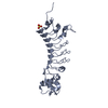
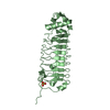
| Entry | Database: PDB / ID: 2bph | ||||||
|---|---|---|---|---|---|---|---|
| Title | STRUCTURE OF MURINE DECTIN-1 | ||||||
 Components Components | DECTIN-1 CLEC7A CLEC7A | ||||||
 Keywords Keywords | RECEPTOR / DECTIN-1 / BETA-GLUCAN / FUNGAL RECOGNITION / C-TYPE LECTIN-LIKE DOMAIN / CTLD / CARBOHYDRATE | ||||||
| Function / homology |  Function and homology information Function and homology information(1->3)-beta-D-glucan immune receptor activity / positive regulation of lymphocyte activation / detection of yeast / detection of molecule of fungal origin / (1->3)-beta-D-glucan binding / cell recognition / detection of fungus / regulation of calcineurin-NFAT signaling cascade / response to molecule of fungal origin / positive regulation of dendritic cell cytokine production ...(1->3)-beta-D-glucan immune receptor activity / positive regulation of lymphocyte activation / detection of yeast / detection of molecule of fungal origin / (1->3)-beta-D-glucan binding / cell recognition / detection of fungus / regulation of calcineurin-NFAT signaling cascade / response to molecule of fungal origin / positive regulation of dendritic cell cytokine production / cell surface pattern recognition receptor signaling pathway / positive regulation of cell maturation / positive regulation of cytokine production involved in immune response / antifungal innate immune response / opsonin binding / positive regulation of interleukin-23 production / positive regulation of stress-activated MAPK cascade / CLEC7A (Dectin-1) signaling / response to yeast / leukocyte activation involved in immune response / cell activation / phagocytosis, recognition / positive regulation of killing of cells of another organism / cellular response to molecule of fungal origin / positive regulation of T-helper 17 type immune response / positive regulation of cytokine production involved in inflammatory response / positive regulation of monocyte chemotactic protein-1 production / regulation of canonical NF-kappaB signal transduction / pattern recognition receptor activity / phagocytosis, engulfment / polysaccharide binding / positive regulation of wound healing / positive regulation of respiratory burst / stimulatory C-type lectin receptor signaling pathway / positive regulation of interleukin-10 production / positive regulation of calcium-mediated signaling / positive regulation of phagocytosis / positive regulation of interleukin-2 production / positive regulation of interleukin-12 production / positive regulation of superoxide anion generation / positive regulation of interleukin-1 beta production / positive regulation of interleukin-8 production / positive regulation of protein-containing complex assembly / positive regulation of DNA-binding transcription factor activity / cell-cell adhesion / positive regulation of interleukin-6 production / positive regulation of nitric oxide biosynthetic process / positive regulation of tumor necrosis factor production / positive regulation of type II interferon production / positive regulation of peptidyl-tyrosine phosphorylation / positive regulation of NF-kappaB transcription factor activity / carbohydrate binding / positive regulation of canonical NF-kappaB signal transduction / positive regulation of cell migration / inflammatory response / external side of plasma membrane / innate immune response / positive regulation of cell population proliferation / positive regulation of gene expression / cell surface / identical protein binding / metal ion binding / plasma membrane / cytoplasmSimilarity search - Function | ||||||
| Biological species |  MUS MUSCULUS (house mouse) MUS MUSCULUS (house mouse) | ||||||
| Method | X-RAY DIFFRACTION / SYNCHROTRON / MOLECULAR REPLACEMENT / Resolution: 2.2 Å | ||||||
 Authors Authors | Brown, J. / O'Callaghan, C.A. / Marshall, A.S.J. / Gilbert, R.J.C. / Siebold, C. / Gordon, S. / Brown, G.D. / Jones, E.Y. | ||||||
 Citation Citation | Journal: Protein Sci. / Year: 2007 Title: Structure of the Fungal Beta-Glucan-Binding Immune Receptor Dectin-1: Implications for Function. Authors: Brown, J. / O'Callaghan, C.A. / Marshall, A.S.J. / Gilbert, R.J.C. / Siebold, C. / Gordon, S. / Brown, G.D. / Jones, E.Y. | ||||||
| History |
| ||||||
| Remark 700 | SHEET DETERMINATION METHOD: AUTHOR PROVIDED. |
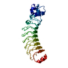
- Structure visualization
| Structure viewer | Molecule: MolmilJmol/JSmol |
|---|
- Downloads & links
-Download
| PDBx/mmCIF format | 2bph.cif.gz | 69.7 KB | Display | PDBx/mmCIF format |
|---|---|---|---|---|
| PDB format | pdb2bph.ent.gz | 50.5 KB | Display | PDB format |
| PDBx/mmJSON format | 2bph.json.gz | Tree view | PDBx/mmJSON format | |
| Others |  Other downloads Other downloads |
-Validation report
| Arichive directory | https://data.pdbj.org/pub/pdb/validation_reports/bp/2bphftp://data.pdbj.org/pub/pdb/validation_reports/bp/2bph | HTTPS FTP |
|---|
-Related structure data
| Related structure data |  2bpdSC  2bpeC  2cl8C S: Starting model for refinement C: citing same article ( |
|---|---|
| Similar structure data |


-Links
 PDBj
PDBj


- Assembly
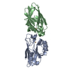
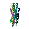
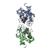
| Deposited unit | 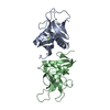
| ||||||||||||||||||
|---|---|---|---|---|---|---|---|---|---|---|---|---|---|---|---|---|---|---|---|
| 1 |
| ||||||||||||||||||
| Unit cell |
| ||||||||||||||||||
| Noncrystallographic symmetry (NCS) | NCS domain:
NCS domain segments: Component-ID: 1 / Ens-ID: 1 / Beg auth comp-ID: GLN / Beg label comp-ID: GLN / End auth comp-ID: LEU / End label comp-ID: LEU / Refine code: 5 / Auth seq-ID: 117 - 244 / Label seq-ID: 6 - 133
NCS oper: (Code: given Matrix: (-0.99869, 0.01789, 0.04796), Vector : |
-Components
| #1: Protein | CLEC7A Mass: 16227.040 Da / Num. of mol.: 2 Fragment: EXTRACELLULAR BETA-GLUCAN RECOGNITION DOMAIN, RESIDUES 113-244 Source method: isolated from a genetically manipulated source Source: (gene. exp.) MUS MUSCULUS (house mouse) / Cell line: RAW264.7 / Plasmid: PET22B / Production host:  ESCHERICHIA COLI (E. coli) / Variant (production host): Rosetta / References: UniProt: Q6QLQ4 ESCHERICHIA COLI (E. coli) / Variant (production host): Rosetta / References: UniProt: Q6QLQ4#2: Chemical |   Mass: 24.305 Da / Num. of mol.: 2 / Source method: obtained synthetically / Formula: Mg Mass: 24.305 Da / Num. of mol.: 2 / Source method: obtained synthetically / Formula: Mg#3: Water | ChemComp-HOH / | Water Mass: 18.015 Da / Num. of mol.: 170 / Source method: isolated from a natural source / Formula: H2O Mass: 18.015 Da / Num. of mol.: 170 / Source method: isolated from a natural source / Formula: H2O |
|---|
-Experimental details
-Experiment
| Experiment | Method: X-RAY DIFFRACTION / Number of used crystals: 1 |
|---|
- Sample preparation
| Crystal | Density Matthews: 2.13 Å3/Da / Density % sol: 42.31 % |
|---|---|
| Crystal grow | pH: 6.5 Details: 0.2 M POTASSIUM CHLORIDE, 0.05M SODIUM CACODYLATE PH6.5, 0.1M MAGNESIUM ACETATE, 10% PEG 8000, pH 6.50 |
-Data collection
| Diffraction | Mean temperature: 100 K |
|---|---|
| Diffraction source | Source: SYNCHROTRON / Site: ESRF  / Beamline: ID14-3 / Wavelength: 0.931 / Beamline: ID14-3 / Wavelength: 0.931 |
| Detector | Type: MARRESEARCH / Detector: CCD / Date: Jul 15, 2004 / Details: TOROIDAL MIRROR |
| Radiation | Monochromator: DIAMOND (111), GE(220) / Protocol: SINGLE WAVELENGTH / Monochromatic (M) / Laue (L): M / Scattering type: x-ray |
| Radiation wavelength | Wavelength: 0.931 Å / Relative weight: 1 |
| Reflection | Resolution: 2.2→30 Å / Num. obs: 13478 / % possible obs: 99.8 % / Observed criterion σ(I): -3 / Redundancy: 4.2 % / Rmerge(I) obs: 0.12 / Net I/σ(I): 12.8 |
| Reflection shell | Resolution: 2.2→2.28 Å / Redundancy: 3.8 % / Rmerge(I) obs: 0.74 / Mean I/σ(I) obs: 2.1 / % possible all: 99.5 |
- Processing
| Software |
| ||||||||||||||||||||||||||||||||||||||||||||||||||||||||||||||||||||||||||||||||||||||||||||||||||||||||||||||||||||||||||||||||||||||||||||||||||||||||||||||||||||||||||||||||||||||
|---|---|---|---|---|---|---|---|---|---|---|---|---|---|---|---|---|---|---|---|---|---|---|---|---|---|---|---|---|---|---|---|---|---|---|---|---|---|---|---|---|---|---|---|---|---|---|---|---|---|---|---|---|---|---|---|---|---|---|---|---|---|---|---|---|---|---|---|---|---|---|---|---|---|---|---|---|---|---|---|---|---|---|---|---|---|---|---|---|---|---|---|---|---|---|---|---|---|---|---|---|---|---|---|---|---|---|---|---|---|---|---|---|---|---|---|---|---|---|---|---|---|---|---|---|---|---|---|---|---|---|---|---|---|---|---|---|---|---|---|---|---|---|---|---|---|---|---|---|---|---|---|---|---|---|---|---|---|---|---|---|---|---|---|---|---|---|---|---|---|---|---|---|---|---|---|---|---|---|---|---|---|---|---|
| Refinement | Method to determine structure: MOLECULAR REPLACEMENT Starting model: PDB ENTRY 2BPD Resolution: 2.2→30 Å / Cor.coef. Fo:Fc: 0.943 / Cor.coef. Fo:Fc free: 0.914 / SU B: 14.208 / SU ML: 0.197 / TLS residual ADP flag: LIKELY RESIDUAL / Cross valid method: THROUGHOUT / ESU R: 0.339 / ESU R Free: 0.239 / Stereochemistry target values: MAXIMUM LIKELIHOOD / Details: HYDROGENS HAVE BEEN ADDED IN THE RIDING POSITIONS.
| ||||||||||||||||||||||||||||||||||||||||||||||||||||||||||||||||||||||||||||||||||||||||||||||||||||||||||||||||||||||||||||||||||||||||||||||||||||||||||||||||||||||||||||||||||||||
| Displacement parameters | Biso mean: 33.59 Å2
| ||||||||||||||||||||||||||||||||||||||||||||||||||||||||||||||||||||||||||||||||||||||||||||||||||||||||||||||||||||||||||||||||||||||||||||||||||||||||||||||||||||||||||||||||||||||
| Refinement step | Cycle: LAST / Resolution: 2.2→30 Å
| ||||||||||||||||||||||||||||||||||||||||||||||||||||||||||||||||||||||||||||||||||||||||||||||||||||||||||||||||||||||||||||||||||||||||||||||||||||||||||||||||||||||||||||||||||||||
| Refine LS restraints |
|