 Movie
Movie Controller


+ Open data

- Basic information
| Entry | Database: PDB / ID: 6fej | ||||||
|---|---|---|---|---|---|---|---|
| Title | Anabaena Apo-C-Terminal Domain Homolog Protein | ||||||
 Components Components | All4940 protein | ||||||
 Keywords Keywords |  PHOTOSYNTHESIS / carotene cyanobacteria photoprotection urea PHOTOSYNTHESIS / carotene cyanobacteria photoprotection urea | ||||||
| Function / homology |  Function and homology information Function and homology informationNuclear transport factor 2 domain / Nuclear transport factor 2 (NTF2) domain / SnoaL-like domain / Nuclear Transport Factor 2; Chain: A, - #50 / NTF2-like domain superfamily / Nuclear Transport Factor 2; Chain: A, / Roll / Alpha Beta Similarity search - Domain/homology | ||||||
| Biological species |  Nostoc sp. PCC 7120 (bacteria) Nostoc sp. PCC 7120 (bacteria) | ||||||
| Method | X-RAY DIFFRACTION / SYNCHROTRON / MOLECULAR REPLACEMENT / Resolution: 2.75 Å | ||||||
 Authors Authors | Harris, D. / Wilson, A. / Muzzopappa, F. / Kirilovsky, D. / Adir, N. | ||||||
| Funding support |  Israel, 1items Israel, 1items
| ||||||
 Citation Citation | Journal: Commun Biol / Year: 2018 Title: Structural rearrangements in the C-terminal domain homolog of Orange Carotenoid Protein are crucial for carotenoid transfer. Authors: Harris, D. / Wilson, A. / Muzzopappa, F. / Sluchanko, N.N. / Friedrich, T. / Maksimov, E.G. / Kirilovsky, D. / Adir, N. | ||||||
| History |
|
- Structure visualization
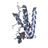
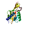
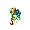
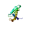
| Structure viewer | Molecule: MolmilJmol/JSmol |
|---|
- Downloads & links
-Download
| PDBx/mmCIF format | 6fej.cif.gz | 107.8 KB | Display | PDBx/mmCIF format |
|---|---|---|---|---|
| PDB format | pdb6fej.ent.gz | 83.5 KB | Display | PDB format |
| PDBx/mmJSON format | 6fej.json.gz | Tree view | PDBx/mmJSON format | |
| Others |  Other downloads Other downloads |
-Validation report
| Arichive directory | https://data.pdbj.org/pub/pdb/validation_reports/fe/6fejftp://data.pdbj.org/pub/pdb/validation_reports/fe/6fej | HTTPS FTP |
|---|
-Related structure data
| Related structure data |  5ui2S S: Starting model for refinement |
|---|---|
| Similar structure data |


-Links
 PDBj
PDBj
- Assembly
| Deposited unit | 
| ||||||||
|---|---|---|---|---|---|---|---|---|---|
| 1 | 
| ||||||||
| 2 | 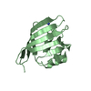
| ||||||||
| 3 | 
| ||||||||
| Unit cell |
|
-Components
| #1: Protein | Mass: 13292.953 Da / Num. of mol.: 2 Source method: isolated from a genetically manipulated source Source: (gene. exp.) Nostoc sp. PCC 7120 (bacteria) / Gene: all4940 / Production host: Escherichia coli (E. coli) / References: UniProt: Q8YMJ3#2: Chemical | Urea  Mass: 60.055 Da / Num. of mol.: 3 / Source method: obtained synthetically / Formula: CH4N2O Mass: 60.055 Da / Num. of mol.: 3 / Source method: obtained synthetically / Formula: CH4N2O#3: Water | ChemComp-HOH / | Water Mass: 18.015 Da / Num. of mol.: 15 / Source method: isolated from a natural source / Formula: H2O Mass: 18.015 Da / Num. of mol.: 15 / Source method: isolated from a natural source / Formula: H2O |
|---|
-Experimental details
-Experiment
| Experiment | Method: X-RAY DIFFRACTION / Number of used crystals: 1 |
|---|
- Sample preparation
| Crystal | Density Matthews: 2.91 Å3/Da / Density % sol: 57.7 % / Description: elongated hexagonal rod |
|---|---|
| Crystal grow | Temperature: 288 K / Method: vapor diffusion, hanging drop / pH: 4.2 / Details: 0.1M citric acid 25% w/v PEG 3350 |
-Data collection
| Diffraction | Mean temperature: 100 K |
|---|---|
| Diffraction source | Source: SYNCHROTRON / Site: ESRF  / Beamline: MASSIF-1 / Wavelength: 0.966 Å / Beamline: MASSIF-1 / Wavelength: 0.966 Å |
| Detector | Type: DECTRIS PILATUS3 2M / Detector: PIXEL / Date: Aug 21, 2017 |
| Radiation | Monochromator: C(110) / Protocol: SINGLE WAVELENGTH / Monochromatic (M) / Laue (L): M / Scattering type: x-ray |
| Radiation wavelength | Wavelength: 0.966 Å / Relative weight: 1 |
| Reflection | Resolution: 2.75→70.22 Å / Num. obs: 8819 / % possible obs: 99.8 % / Redundancy: 5.6 % / CC1/2: 0.935 / Rmerge(I) obs: 0.071 / Rpim(I) all: 0.032 / Net I/σ(I): 11.7 |
| Reflection shell | Resolution: 2.75→2.848 Å / Rmerge(I) obs: 0.334 / Mean I/σ(I) obs: 5 / Num. unique obs: 1247 / CC1/2: 0.898 / Rpim(I) all: 0.142 / % possible all: 100 |
- Processing
| Software |
| |||||||||||||||||||||||||||||||||||||||||||||||||||||||||||||||||||||||||||
|---|---|---|---|---|---|---|---|---|---|---|---|---|---|---|---|---|---|---|---|---|---|---|---|---|---|---|---|---|---|---|---|---|---|---|---|---|---|---|---|---|---|---|---|---|---|---|---|---|---|---|---|---|---|---|---|---|---|---|---|---|---|---|---|---|---|---|---|---|---|---|---|---|---|---|---|---|
| Refinement | Method to determine structure: MOLECULAR REPLACEMENT Starting model: 5UI2 Resolution: 2.75→70.22 Å / Cor.coef. Fo:Fc: 0.943 / Cor.coef. Fo:Fc free: 0.898 / Cross valid method: THROUGHOUT / σ(F): 0 / ESU R Free: 0.408 Details: HYDROGENS HAVE BEEN ADDED IN THE RIDING POSITIONS U VALUES : WITH TLS ADDED
| |||||||||||||||||||||||||||||||||||||||||||||||||||||||||||||||||||||||||||
| Solvent computation | Ion probe radii: 0.8 Å / Shrinkage radii: 0.8 Å / VDW probe radii: 1.2 Å | |||||||||||||||||||||||||||||||||||||||||||||||||||||||||||||||||||||||||||
| Displacement parameters | Biso max: 119.62 Å2 / Biso mean: 67.309 Å2 / Biso min: 22.71 Å2
| |||||||||||||||||||||||||||||||||||||||||||||||||||||||||||||||||||||||||||
| Refinement step | Cycle: final / Resolution: 2.75→70.22 Å
| |||||||||||||||||||||||||||||||||||||||||||||||||||||||||||||||||||||||||||
| LS refinement shell | Resolution: 2.75→2.821 Å / Rfactor Rfree error: 0 / Total num. of bins used: 20
| |||||||||||||||||||||||||||||||||||||||||||||||||||||||||||||||||||||||||||
| Refinement TLS params. | Method: refined / Refine-ID: X-RAY DIFFRACTION
| |||||||||||||||||||||||||||||||||||||||||||||||||||||||||||||||||||||||||||
| Refinement TLS group |
|
