 Movie
Movie Controller

[English] 日本語
 Yorodumi
- PDB-2bnm: The structure of Hydroxypropylphosphonic acid epoxidase from S. w... -
+ Open data

- Basic information
| Entry | Database: PDB / ID: 2bnm | ||||||
|---|---|---|---|---|---|---|---|
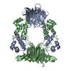
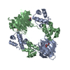
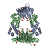
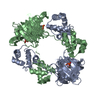
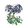
| Title | The structure of Hydroxypropylphosphonic acid epoxidase from S. wedmorenis. | ||||||
 Components Components | EPOXIDASE | ||||||
 Keywords Keywords |  OXIDOREDUCTASE / EPOXIDASE / CUPIN / HTH / CATION-DEPENDANT / ZINC / FOSFOMYCIN OXIDOREDUCTASE / EPOXIDASE / CUPIN / HTH / CATION-DEPENDANT / ZINC / FOSFOMYCIN | ||||||
| Function / homology |  Function and homology information(S)-2-hydroxypropylphosphonic acid epoxidase / phosphinothricin biosynthetic process / oxidoreductase activity, acting on paired donors, with oxidation of a pair of donors resulting in the reduction of molecular oxygen to two molecules of water / dioxygenase activity / antibiotic biosynthetic process / ferrous iron binding / protein homotetramerization / DNA binding Function and homology information(S)-2-hydroxypropylphosphonic acid epoxidase / phosphinothricin biosynthetic process / oxidoreductase activity, acting on paired donors, with oxidation of a pair of donors resulting in the reduction of molecular oxygen to two molecules of water / dioxygenase activity / antibiotic biosynthetic process / ferrous iron binding / protein homotetramerization / DNA bindingSimilarity search - Function | ||||||
| Biological species |  STREPTOMYCES WEDMORENSIS (bacteria) STREPTOMYCES WEDMORENSIS (bacteria) | ||||||
| Method | X-RAY DIFFRACTION / SYNCHROTRON / OTHER / Resolution: 1.7 Å | ||||||
 Authors Authors | McLuskey, K. / Cameron, S. / Hunter, W.N. | ||||||
 Citation Citation | Journal: Proc.Natl.Acad.Sci.USA / Year: 2005 Title: Structure and Reactivity of Hydroxypropylphosphonic Acid Epoxidase in Fosfomycin Biosynthesis by a Cation- and Flavin-Dependent Mechanism. Authors: Mcluskey, K. / Cameron, S. / Hammerschmidt, F. / Hunter, W.N. #1: Journal: Acta Crystallogr.,Sect.F / Year: 2005 Title: Initiating a Crystallographic Analysis of Recombinant (S)-2-Hydroxypropylphosphonic Acid Epoxidase from Streptomyces Wedmorensis. Authors: Cameron, S. / Mcluskey, K. / Chamberlayne, R. / Hallyburton, I. / Hunter, W.N. | ||||||
| History |
|
- Structure visualization
| Structure viewer | Molecule: MolmilJmol/JSmol |
|---|
- Downloads & links
-Download
| PDBx/mmCIF format | 2bnm.cif.gz | 101.6 KB | Display | PDBx/mmCIF format |
|---|---|---|---|---|
| PDB format | pdb2bnm.ent.gz | 83.6 KB | Display | PDB format |
| PDBx/mmJSON format | 2bnm.json.gz | Tree view | PDBx/mmJSON format | |
| Others |  Other downloads Other downloads |
-Validation report
| Arichive directory | https://data.pdbj.org/pub/pdb/validation_reports/bn/2bnmftp://data.pdbj.org/pub/pdb/validation_reports/bn/2bnm | HTTPS FTP |
|---|
-Related structure data


-Links
 PDBj
PDBj


- Assembly
| Deposited unit | 
| ||||||||
|---|---|---|---|---|---|---|---|---|---|
| 1 | 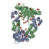
| ||||||||
| Unit cell |
| ||||||||
| Noncrystallographic symmetry (NCS) | NCS oper: (Code: given Matrix: (-0.99993, 0.00565, -0.01069), Vector : |
-Components
| #1: Protein | Mass: 21361.127 Da / Num. of mol.: 2 Source method: isolated from a genetically manipulated source Source: (gene. exp.) STREPTOMYCES WEDMORENSIS (bacteria) / Plasmid: PET15B / Production host: ESCHERICHIA COLI (E. coli) / Strain (production host): BL21(DE3) / References: UniProt: Q56185#2: Chemical |   Mass: 65.409 Da / Num. of mol.: 2 / Source method: obtained synthetically / Formula: Zn Mass: 65.409 Da / Num. of mol.: 2 / Source method: obtained synthetically / Formula: Zn#3: Chemical | ChemComp-SO4 / Sulfate  Mass: 96.063 Da / Num. of mol.: 15 / Source method: obtained synthetically / Formula: SO4 Mass: 96.063 Da / Num. of mol.: 15 / Source method: obtained synthetically / Formula: SO4#4: Water | ChemComp-HOH / | Water Mass: 18.015 Da / Num. of mol.: 547 / Source method: isolated from a natural source / Formula: H2O Mass: 18.015 Da / Num. of mol.: 547 / Source method: isolated from a natural source / Formula: H2O |
|---|
-Experimental details
-Experiment
| Experiment | Method: X-RAY DIFFRACTION / Number of used crystals: 1 |
|---|
- Sample preparation
| Crystal | Density Matthews: 2.27 Å3/Da / Density % sol: 45.36 % Description: MODEL USED FOR RIGID BODY REFINEMENT HAS JUST BEEN DEPOSITED. |
|---|---|
| Crystal grow | pH: 7.5 / Details: 2.1 M AMMONIUM SULPHATE, 100 MM TRIS-HCL PH 7.5 |
-Data collection
| Diffraction | Mean temperature: 100 K |
|---|---|
| Diffraction source | Source: SYNCHROTRON / Site: ESRF  / Beamline: BM14 / Wavelength: 1.27 / Beamline: BM14 / Wavelength: 1.27 |
| Detector | Type: MARRESEARCH / Detector: CCD / Date: Apr 20, 2004 |
| Radiation | Protocol: SINGLE WAVELENGTH / Monochromatic (M) / Laue (L): M / Scattering type: x-ray |
| Radiation wavelength | Wavelength: 1.27 Å / Relative weight: 1 |
| Reflection | Resolution: 1.7→30 Å / Num. obs: 53414 / % possible obs: 98.5 % / Observed criterion σ(I): 2 / Redundancy: 11.8 % / Rmerge(I) obs: 0.05 / Net I/σ(I): 41.5 |
| Reflection shell | Resolution: 1.7→1.76 Å / Rmerge(I) obs: 0.27 / Mean I/σ(I) obs: 4.9 / % possible all: 82.7 |
- Processing
| Software |
| ||||||||||||||||||||||||||||||||||||||||||||||||||||||||||||||||||||||||||||||||||||||||||||||||||||||||||||||||||||||||||||||||||||||||||||||||||||||||||||||||||||||||||||||||||||||
|---|---|---|---|---|---|---|---|---|---|---|---|---|---|---|---|---|---|---|---|---|---|---|---|---|---|---|---|---|---|---|---|---|---|---|---|---|---|---|---|---|---|---|---|---|---|---|---|---|---|---|---|---|---|---|---|---|---|---|---|---|---|---|---|---|---|---|---|---|---|---|---|---|---|---|---|---|---|---|---|---|---|---|---|---|---|---|---|---|---|---|---|---|---|---|---|---|---|---|---|---|---|---|---|---|---|---|---|---|---|---|---|---|---|---|---|---|---|---|---|---|---|---|---|---|---|---|---|---|---|---|---|---|---|---|---|---|---|---|---|---|---|---|---|---|---|---|---|---|---|---|---|---|---|---|---|---|---|---|---|---|---|---|---|---|---|---|---|---|---|---|---|---|---|---|---|---|---|---|---|---|---|---|---|
| Refinement | Method to determine structure: OTHER / Resolution: 1.7→30 Å / Cor.coef. Fo:Fc: 0.96 / Cor.coef. Fo:Fc free: 0.945 / SU B: 2.055 / SU ML: 0.069 / Cross valid method: THROUGHOUT / ESU R: 0.107 / ESU R Free: 0.11 / Stereochemistry target values: MAXIMUM LIKELIHOOD / Details: HYDROGENS HAVE BEEN ADDED IN THE RIDING POSITIONS.
| ||||||||||||||||||||||||||||||||||||||||||||||||||||||||||||||||||||||||||||||||||||||||||||||||||||||||||||||||||||||||||||||||||||||||||||||||||||||||||||||||||||||||||||||||||||||
| Solvent computation | Ion probe radii: 0.8 Å / Shrinkage radii: 0.8 Å / VDW probe radii: 1.2 Å / Solvent model: BABINET MODEL WITH MASK | ||||||||||||||||||||||||||||||||||||||||||||||||||||||||||||||||||||||||||||||||||||||||||||||||||||||||||||||||||||||||||||||||||||||||||||||||||||||||||||||||||||||||||||||||||||||
| Displacement parameters | Biso mean: 27.73 Å2
| ||||||||||||||||||||||||||||||||||||||||||||||||||||||||||||||||||||||||||||||||||||||||||||||||||||||||||||||||||||||||||||||||||||||||||||||||||||||||||||||||||||||||||||||||||||||
| Refinement step | Cycle: LAST / Resolution: 1.7→30 Å
| ||||||||||||||||||||||||||||||||||||||||||||||||||||||||||||||||||||||||||||||||||||||||||||||||||||||||||||||||||||||||||||||||||||||||||||||||||||||||||||||||||||||||||||||||||||||
| Refine LS restraints |
|