 Movie
Movie Controller

[English] 日本語
 Yorodumi
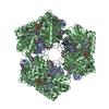
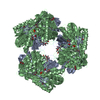
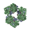
- PDB-2bjk: Crystal Analysis of 1-Pyrroline-5-Carboxylate Dehydrogenase from ... -
+ Open data

- Basic information
| Entry | Database: PDB / ID: 2bjk | ||||||
|---|---|---|---|---|---|---|---|
| Title | Crystal Analysis of 1-Pyrroline-5-Carboxylate Dehydrogenase from Thermus with bound NAD and citrate. | ||||||
 Components Components | 1-PYRROLINE-5-CARBOXYLATE DEHYDROGENASE | ||||||
 Keywords Keywords | OXIDOREDUCTASE / 1-PYRROLINE-5-CARBOXYLATE / DEHYROGENASE | ||||||
| Function / homology |  Function and homology information1-pyrroline-5-carboxylate dehydrogenase activity / L-glutamate gamma-semialdehyde dehydrogenase / proline catabolic process to glutamate / cytoplasmic side of plasma membrane / nucleotide binding Function and homology information1-pyrroline-5-carboxylate dehydrogenase activity / L-glutamate gamma-semialdehyde dehydrogenase / proline catabolic process to glutamate / cytoplasmic side of plasma membrane / nucleotide bindingSimilarity search - Function | ||||||
| Biological species |   THERMUS THERMOPHILUS (bacteria) THERMUS THERMOPHILUS (bacteria) | ||||||
| Method | X-RAY DIFFRACTION / SYNCHROTRON / MOLECULAR REPLACEMENT / Resolution: 1.4 Å | ||||||
 Authors Authors | Inagaki, E. / Tahirov, T.H. | ||||||
 Citation Citation | Journal: J.Mol.Biol. / Year: 2006 Title: Crystal Structure of Thermus Thermophilus Delta(1)- Pyrroline-5-Carboxylate Dehydrogenase. Authors: Inagaki, E. / Ohshima, N. / Takahashi, H. / Kuroishi, C. / Yokoyama, S. / Tahirov, T.H. | ||||||
| History |
|
- Structure visualization
| Structure viewer | Molecule: MolmilJmol/JSmol |
|---|
- Downloads & links
-Download
| PDBx/mmCIF format | 2bjk.cif.gz | 245.8 KB | Display | PDBx/mmCIF format |
|---|---|---|---|---|
| PDB format | pdb2bjk.ent.gz | 197.3 KB | Display | PDB format |
| PDBx/mmJSON format | 2bjk.json.gz | Tree view | PDBx/mmJSON format | |
| Others |  Other downloads Other downloads |
-Validation report
| Arichive directory | https://data.pdbj.org/pub/pdb/validation_reports/bj/2bjkftp://data.pdbj.org/pub/pdb/validation_reports/bj/2bjk | HTTPS FTP |
|---|
-Related structure data
| Related structure data |  1uzbSC 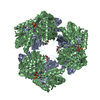 2bhpC 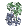 2bhqC 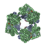 2bjaC 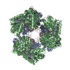 2iy6C C: citing same article ( S: Starting model for refinement |
|---|---|
| Similar structure data |


-Links
 PDBj
PDBj


- Assembly
| Deposited unit | 
| ||||||||
|---|---|---|---|---|---|---|---|---|---|
| 1 | 
| ||||||||
| Unit cell |
| ||||||||
| Noncrystallographic symmetry (NCS) | NCS oper: (Code: given Matrix: (0.97687, -0.21369, -0.00718), Vector : |
-Components
-Protein , 1 types, 2 molecules AB
| #1: Protein | Mass: 57113.906 Da / Num. of mol.: 2 Source method: isolated from a genetically manipulated source Source: (gene. exp.) THERMUS THERMOPHILUS (bacteria) / Strain: HB8 / Plasmid: PET11A / Production host: ESCHERICHIA COLI (E. coli) / Strain (production host): BL21(DE3) / References: UniProt: Q5SI02, EC: 1.5.1.12 |
|---|
-Non-polymers , 6 types, 1376 molecules 


| #2: Chemical | Nicotinamide adenine dinucleotide Mass: 663.425 Da / Num. of mol.: 2 / Source method: obtained synthetically / Formula: C21H27N7O14P2 / Comment: NAD*YM Mass: 663.425 Da / Num. of mol.: 2 / Source method: obtained synthetically / Formula: C21H27N7O14P2 / Comment: NAD*YM#3: Chemical | Citric acid Mass: 189.100 Da / Num. of mol.: 2 / Source method: obtained synthetically / Formula: C6H5O7 Mass: 189.100 Da / Num. of mol.: 2 / Source method: obtained synthetically / Formula: C6H5O7#4: Chemical | ChemComp-MRD / ( 2-Methyl-2,4-pentanediol Mass: 118.174 Da / Num. of mol.: 4 / Source method: obtained synthetically / Formula: C6H14O2 / Comment: precipitant*YM Mass: 118.174 Da / Num. of mol.: 4 / Source method: obtained synthetically / Formula: C6H14O2 / Comment: precipitant*YM#5: Chemical | ChemComp-MPD / ( 2-Methyl-2,4-pentanediol Mass: 118.174 Da / Num. of mol.: 5 / Source method: obtained synthetically / Formula: C6H14O2 / Comment: precipitant*YM Mass: 118.174 Da / Num. of mol.: 5 / Source method: obtained synthetically / Formula: C6H14O2 / Comment: precipitant*YM#6: Chemical | Chloride Mass: 35.453 Da / Num. of mol.: 2 / Source method: obtained synthetically / Formula: Cl Mass: 35.453 Da / Num. of mol.: 2 / Source method: obtained synthetically / Formula: Cl#7: Water | ChemComp-HOH / | WaterMass: 18.015 Da / Num. of mol.: 1361 / Source method: isolated from a natural source / Formula: H2O |
|---|
-Experimental details
-Experiment
| Experiment | Method: X-RAY DIFFRACTION / Number of used crystals: 1 |
|---|
- Sample preparation
| Crystal | Density Matthews: 2.5 Å3/Da / Density % sol: 50.1 % |
|---|---|
| Crystal grow | pH: 5.2 Details: PROTEIN WAS CRYSTALLIZED FROM 32% MPD, 50 MM SODIUM CITRATE/HCL, PH 5.2; THEN SOAKED IN 1 MM NAD AND 10MM MGCL2. |
-Data collection
| Diffraction | Mean temperature: 100 K |
|---|---|
| Diffraction source | Source: SYNCHROTRON / Site: SPring-8  / Beamline: BL26B1 / Wavelength: 0.8 / Beamline: BL26B1 / Wavelength: 0.8 |
| Detector | Type: RIGAKU RAXIS-V / Detector: IMAGE PLATE / Date: Oct 22, 2002 / Details: MIRRORS |
| Radiation | Monochromator: SILICON CRYSTAL / Protocol: SINGLE WAVELENGTH / Monochromatic (M) / Laue (L): M / Scattering type: x-ray |
| Radiation wavelength | Wavelength: 0.8 Å / Relative weight: 1 |
| Reflection | Resolution: 1.04→50 Å / Num. obs: 511404 / % possible obs: 97.6 % / Observed criterion σ(I): -0.5 / Redundancy: 2.5 % / Biso Wilson estimate: 12 Å2 / Rmerge(I) obs: 0.07 / Net I/σ(I): 16.9 |
| Reflection shell | Resolution: 1.04→1.05 Å / Redundancy: 2.1 % / Rmerge(I) obs: 0.59 / Mean I/σ(I) obs: 1.7 / % possible all: 94 |
- Processing
| Software |
| ||||||||||||||||||||||||||||||||||||||||||||||||||||||||||||||||||||||||||||||||
|---|---|---|---|---|---|---|---|---|---|---|---|---|---|---|---|---|---|---|---|---|---|---|---|---|---|---|---|---|---|---|---|---|---|---|---|---|---|---|---|---|---|---|---|---|---|---|---|---|---|---|---|---|---|---|---|---|---|---|---|---|---|---|---|---|---|---|---|---|---|---|---|---|---|---|---|---|---|---|---|---|---|
| Refinement | Method to determine structure: MOLECULAR REPLACEMENT Starting model: PDB ENTRY 1UZB Resolution: 1.4→33.29 Å / Rfactor Rfree error: 0.002 / Data cutoff high absF: 1404023.09 / Isotropic thermal model: RESTRAINED / Cross valid method: THROUGHOUT / σ(F): 0
| ||||||||||||||||||||||||||||||||||||||||||||||||||||||||||||||||||||||||||||||||
| Solvent computation | Solvent model: FLAT MODEL / Bsol: 44.5273 Å2 / ksol: 0.32476 e/Å3 | ||||||||||||||||||||||||||||||||||||||||||||||||||||||||||||||||||||||||||||||||
| Displacement parameters | Biso mean: 13.9 Å2
| ||||||||||||||||||||||||||||||||||||||||||||||||||||||||||||||||||||||||||||||||
| Refine analyze |
| ||||||||||||||||||||||||||||||||||||||||||||||||||||||||||||||||||||||||||||||||
| Refinement step | Cycle: LAST / Resolution: 1.4→33.29 Å
| ||||||||||||||||||||||||||||||||||||||||||||||||||||||||||||||||||||||||||||||||
| Refine LS restraints |
| ||||||||||||||||||||||||||||||||||||||||||||||||||||||||||||||||||||||||||||||||
| LS refinement shell | Resolution: 1.4→1.49 Å / Rfactor Rfree error: 0.006 / Total num. of bins used: 6
| ||||||||||||||||||||||||||||||||||||||||||||||||||||||||||||||||||||||||||||||||
| Xplor file |
|
