 Movie
Movie Controller


+ Open data

- Basic information
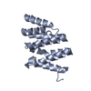
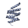
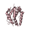
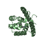
| Entry | Database: PDB / ID: 2bf0 | ||||||
|---|---|---|---|---|---|---|---|
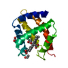
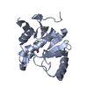
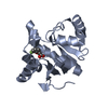
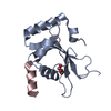
| Title | crystal structure of the rpr of pcf11 | ||||||
 Components Components | PCF11 | ||||||
 Keywords Keywords |  TRANSCRIPTION / RNA / PHOSPHORYLATION TRANSCRIPTION / RNA / PHOSPHORYLATION | ||||||
| Function / homology |  Function and homology information Function and homology informationtermination of RNA polymerase II transcription, poly(A)-coupled / termination of RNA polymerase II transcription, exosome-dependent / mRNA cleavage factor complex / termination of RNA polymerase II transcription / : / RNA polymerase II complex binding / mRNA binding / nucleus / cytosol / cytoplasmSimilarity search - Function | ||||||
| Biological species |  SACCHAROMYCES CEREVISIAE (brewer's yeast) SACCHAROMYCES CEREVISIAE (brewer's yeast) | ||||||
| Method | X-RAY DIFFRACTION / SYNCHROTRON / MAD / Resolution: 2.3 Å | ||||||
 Authors Authors | Noble, C.G. / Hollingworth, D. / Martin, S.R. / Adeniran, V.E. / Smerdon, S.J. / Kelly, G. / Taylor, I.A. / Ramos, A. | ||||||
 Citation Citation | Journal: Nat.Struct.Mol.Biol. / Year: 2005 Title: Key Features of the Interaction between Pcf11 Cid and RNA Polymerase II Ctd. Authors: Noble, C.G. / Hollingworth, D. / Martin, S.R. / Adeniran, V.E. / Smerdon, S.J. / Kelly, G. / Taylor, I.A. / Ramos, A. | ||||||
| History |
|
- Structure visualization
| Structure viewer | Molecule: MolmilJmol/JSmol |
|---|
- Downloads & links
-Download
| PDBx/mmCIF format | 2bf0.cif.gz | 37.1 KB | Display | PDBx/mmCIF format |
|---|---|---|---|---|
| PDB format | pdb2bf0.ent.gz | 29.2 KB | Display | PDB format |
| PDBx/mmJSON format | 2bf0.json.gz | Tree view | PDBx/mmJSON format | |
| Others |  Other downloads Other downloads |
-Validation report
| Arichive directory | https://data.pdbj.org/pub/pdb/validation_reports/bf/2bf0ftp://data.pdbj.org/pub/pdb/validation_reports/bf/2bf0 | HTTPS FTP |
|---|
-Related structure data
| Related structure data | |
|---|---|
| Similar structure data |


-Links
 PDBj
PDBj
- Assembly
| Deposited unit | 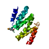
| ||||||||
|---|---|---|---|---|---|---|---|---|---|
| 1 |
| ||||||||
| Unit cell |
|
-Components
| #1: Protein | Mass: 16633.701 Da / Num. of mol.: 1 / Fragment: CID, RESIDUES 1-138 Source method: isolated from a genetically manipulated source Source: (gene. exp.) SACCHAROMYCES CEREVISIAE (brewer's yeast)Plasmid: PGEX6P1 / Production host:  ESCHERICHIA COLI (E. coli) / Strain (production host): BL21(DE3) / References: UniProt: P39081 ESCHERICHIA COLI (E. coli) / Strain (production host): BL21(DE3) / References: UniProt: P39081 |
|---|---|
| #2: Chemical | ChemComp-CA /   Mass: 40.078 Da / Num. of mol.: 1 / Source method: obtained synthetically / Formula: Ca Mass: 40.078 Da / Num. of mol.: 1 / Source method: obtained synthetically / Formula: Ca |
| #3: Water | ChemComp-HOH / Water Mass: 18.015 Da / Num. of mol.: 56 / Source method: isolated from a natural source / Formula: H2O Mass: 18.015 Da / Num. of mol.: 56 / Source method: isolated from a natural source / Formula: H2O |
-Experimental details
-Experiment
| Experiment | Method: X-RAY DIFFRACTION / Number of used crystals: 1 |
|---|
- Sample preparation
| Crystal | Density Matthews: 2.4 Å3/Da / Density % sol: 44 % |
|---|---|
| Crystal grow | pH: 7.8 Details: 25% PEG 4000, 0.1 M TRIS PH 7.0, 0.2 M (NH4)2SO4 AND 10 MM CACL2 |
-Data collection
| Diffraction | Mean temperature: 100 K | ||||||||||||
|---|---|---|---|---|---|---|---|---|---|---|---|---|---|
| Diffraction source | Source: SYNCHROTRON / Site: ESRF  / Beamline: ID29 / Wavelength: 0.976,0.9792,0.9795 / Beamline: ID29 / Wavelength: 0.976,0.9792,0.9795 | ||||||||||||
| Detector | Type: ADSC CCD / Detector: CCD / Date: May 15, 2004 | ||||||||||||
| Radiation | Protocol: MAD / Monochromatic (M) / Laue (L): M / Scattering type: x-ray | ||||||||||||
| Radiation wavelength |
| ||||||||||||
| Reflection | Resolution: 2.3→20 Å / Num. obs: 7191 / % possible obs: 100 % / Observed criterion σ(I): 2 / Redundancy: 7 % / Rmerge(I) obs: 0.06 / Net I/σ(I): 36 |
- Processing
| Software |
| ||||||||||||||||||||||||||||||||||||||||||||||||||||||||||||||||||||||||||||||||||||||||||||||||||||||||||||||||||||||||||||||||||||||||||||||||||||||||||||||||||||||||||||||||||||||
|---|---|---|---|---|---|---|---|---|---|---|---|---|---|---|---|---|---|---|---|---|---|---|---|---|---|---|---|---|---|---|---|---|---|---|---|---|---|---|---|---|---|---|---|---|---|---|---|---|---|---|---|---|---|---|---|---|---|---|---|---|---|---|---|---|---|---|---|---|---|---|---|---|---|---|---|---|---|---|---|---|---|---|---|---|---|---|---|---|---|---|---|---|---|---|---|---|---|---|---|---|---|---|---|---|---|---|---|---|---|---|---|---|---|---|---|---|---|---|---|---|---|---|---|---|---|---|---|---|---|---|---|---|---|---|---|---|---|---|---|---|---|---|---|---|---|---|---|---|---|---|---|---|---|---|---|---|---|---|---|---|---|---|---|---|---|---|---|---|---|---|---|---|---|---|---|---|---|---|---|---|---|---|---|
| Refinement | Method to determine structure: MAD / Resolution: 2.3→15 Å / Cor.coef. Fo:Fc: 0.899 / Cor.coef. Fo:Fc free: 0.875 / Cross valid method: THROUGHOUT / ESU R: 0.394 / ESU R Free: 0.262 / Stereochemistry target values: MAXIMUM LIKELIHOOD / Details: HYDROGENS HAVE BEEN ADDED IN THE RIDING POSITIONS.
| ||||||||||||||||||||||||||||||||||||||||||||||||||||||||||||||||||||||||||||||||||||||||||||||||||||||||||||||||||||||||||||||||||||||||||||||||||||||||||||||||||||||||||||||||||||||
| Solvent computation | Ion probe radii: 0.8 Å / Shrinkage radii: 0.8 Å / VDW probe radii: 1.4 Å / Solvent model: BABINET MODEL WITH MASK | ||||||||||||||||||||||||||||||||||||||||||||||||||||||||||||||||||||||||||||||||||||||||||||||||||||||||||||||||||||||||||||||||||||||||||||||||||||||||||||||||||||||||||||||||||||||
| Displacement parameters | Biso mean: 30.88 Å2
| ||||||||||||||||||||||||||||||||||||||||||||||||||||||||||||||||||||||||||||||||||||||||||||||||||||||||||||||||||||||||||||||||||||||||||||||||||||||||||||||||||||||||||||||||||||||
| Refinement step | Cycle: LAST / Resolution: 2.3→15 Å
| ||||||||||||||||||||||||||||||||||||||||||||||||||||||||||||||||||||||||||||||||||||||||||||||||||||||||||||||||||||||||||||||||||||||||||||||||||||||||||||||||||||||||||||||||||||||
| Refine LS restraints |
|