 Movie
Movie Controller

[English] 日本語
 Yorodumi
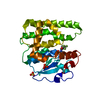
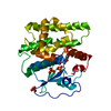
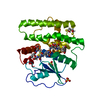
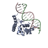
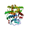
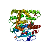
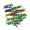
- PDB-2ayb: Crystal structure of HPV6a E2 DNA Binding Domain bound to a 16 ba... -
+ Open data

- Basic information
| Entry | Database: PDB / ID: 2ayb | ||||||
|---|---|---|---|---|---|---|---|
| Title | Crystal structure of HPV6a E2 DNA Binding Domain bound to a 16 base pair DNA target | ||||||
 Components Components |
| ||||||
 Keywords Keywords | TRANSCRIPTION/DNA /  Protein-DNA complex / double helix / beta barrel / TRANSCRIPTION-DNA COMPLEX Protein-DNA complex / double helix / beta barrel / TRANSCRIPTION-DNA COMPLEX | ||||||
| Function / homology |  Function and homology information Function and homology informationviral DNA genome replication / regulation of DNA replication / DNA replication / DNA-binding transcription factor activity / nucleotide binding / DNA-templated transcription / host cell nucleus / DNA bindingSimilarity search - Function | ||||||
| Biological species |  Human papillomavirus type 6a Human papillomavirus type 6a | ||||||
| Method | X-RAY DIFFRACTION / MOLECULAR REPLACEMENT / Resolution: 3.2 Å | ||||||
 Authors Authors | Hooley, E. / Brady, R.L. / Gaston, K. | ||||||
 Citation Citation | Journal: Nucleic Acids Res. / Year: 2006 Title: The recognition of local DNA conformation by the human papillomavirus type 6 E2 protein. Authors: Hooley, E. / Fairweather, V. / Clarke, A.R. / Gaston, K. / Brady, R.L. | ||||||
| History |
|
- Structure visualization
| Structure viewer | Molecule: MolmilJmol/JSmol |
|---|
- Downloads & links
-Download
| PDBx/mmCIF format | 2ayb.cif.gz | 69.1 KB | Display | PDBx/mmCIF format |
|---|---|---|---|---|
| PDB format | pdb2ayb.ent.gz | 48.1 KB | Display | PDB format |
| PDBx/mmJSON format | 2ayb.json.gz | Tree view | PDBx/mmJSON format | |
| Others |  Other downloads Other downloads |
-Validation report
| Arichive directory | https://data.pdbj.org/pub/pdb/validation_reports/ay/2aybftp://data.pdbj.org/pub/pdb/validation_reports/ay/2ayb | HTTPS FTP |
|---|
-Related structure data
| Related structure data | 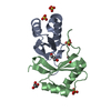 2ayeC 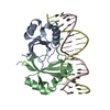 2aygC 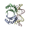 1jj4S  1r8hS C: citing same article ( S: Starting model for refinement |
|---|---|
| Similar structure data |


-Links
 PDBj
PDBj


- Assembly
| Deposited unit | 
| ||||||||||||||||||
|---|---|---|---|---|---|---|---|---|---|---|---|---|---|---|---|---|---|---|---|
| 1 |
| ||||||||||||||||||
| Unit cell |
| ||||||||||||||||||
| Noncrystallographic symmetry (NCS) | NCS domain:
NCS domain segments: Component-ID: 1 / Ens-ID: 1 / Beg auth comp-ID: ALA / Beg label comp-ID: ALA / End auth comp-ID: HIS / End label comp-ID: HIS / Refine code: 1 / Auth seq-ID: 283 - 364 / Label seq-ID: 3 - 85
| ||||||||||||||||||
| Details | The biological assembly is a protein dimer bound to double stranded DNA |
-Components
| #1: DNA chain | Mass: 4898.191 Da / Num. of mol.: 2 / Source method: obtained synthetically #2: Protein | Mass: 10240.854 Da / Num. of mol.: 2 / Fragment: C Terminal Domain Source method: isolated from a genetically manipulated source Source: (gene. exp.) Human papillomavirus type 6a / Genus: Alphapapillomavirus / Species: Human papillomavirus - 6 / Gene: E2 / Plasmid: pKK223-3 / Production host:  Escherichia coli (E. coli) / Strain (production host): Xl1-blue / References: UniProt: Q84294 Escherichia coli (E. coli) / Strain (production host): Xl1-blue / References: UniProt: Q84294#3: Water | ChemComp-HOH / | Water Mass: 18.015 Da / Num. of mol.: 12 / Source method: isolated from a natural source / Formula: H2O Mass: 18.015 Da / Num. of mol.: 12 / Source method: isolated from a natural source / Formula: H2O |
|---|
-Experimental details
-Experiment
| Experiment | Method: X-RAY DIFFRACTION / Number of used crystals: 1 |
|---|
- Sample preparation
| Crystal | Density Matthews: 2.6 Å3/Da / Density % sol: 53 % | ||||||||||||||||||||
|---|---|---|---|---|---|---|---|---|---|---|---|---|---|---|---|---|---|---|---|---|---|
| Crystal grow | Temperature: 291 K / Method: vapor diffusion, hanging drop / pH: 6.5 Details: tri-Sodium citrate dihydrate, pH 6.5, VAPOR DIFFUSION, HANGING DROP, temperature 291K | ||||||||||||||||||||
| Components of the solutions |
|
-Data collection
| Diffraction | Mean temperature: 100 K |
|---|---|
| Diffraction source | Source: ROTATING ANODE / Type: BRUKER X8 PROTEUM / Wavelength: 1.548 Å |
| Detector | Type: BRUKER SMART 6000 / Detector: CCD / Date: May 20, 2005 / Details: mirrors |
| Radiation | Monochromator: Ni Filter / Protocol: SINGLE WAVELENGTH / Monochromatic (M) / Laue (L): M / Scattering type: x-ray |
| Radiation wavelength | Wavelength: 1.548 Å / Relative weight: 1 |
| Reflection | Resolution: 3.2→65.23 Å / Num. all: 5168 / Num. obs: 4928 / % possible obs: 99.37 % / Observed criterion σ(F): 2 / Observed criterion σ(I): 2 / Redundancy: 22.5 % / Rmerge(I) obs: 0.138 / Net I/σ(I): 19.8 |
| Reflection shell | Resolution: 3.2→3.28 Å / Redundancy: 22.5 % / Rmerge(I) obs: 0.387 / Mean I/σ(I) obs: 5.2 / Num. unique all: 362 / % possible all: 100 |
- Processing
| Software |
| ||||||||||||||||||||||||||||||||||||||||||||||||||||||||||||||||||||||||||||||||||||||||||
|---|---|---|---|---|---|---|---|---|---|---|---|---|---|---|---|---|---|---|---|---|---|---|---|---|---|---|---|---|---|---|---|---|---|---|---|---|---|---|---|---|---|---|---|---|---|---|---|---|---|---|---|---|---|---|---|---|---|---|---|---|---|---|---|---|---|---|---|---|---|---|---|---|---|---|---|---|---|---|---|---|---|---|---|---|---|---|---|---|---|---|---|
| Refinement | Method to determine structure: MOLECULAR REPLACEMENT Starting model: Protein of PDB entry 1R8H DNA of PDB entry 1JJ4 Resolution: 3.2→65.23 Å / Cor.coef. Fo:Fc: 0.942 / Cor.coef. Fo:Fc free: 0.843 / SU B: 33.517 / SU ML: 0.402 / TLS residual ADP flag: LIKELY RESIDUAL / Cross valid method: THROUGHOUT / σ(F): 0 / ESU R Free: 0.632 / Stereochemistry target values: MAXIMUM LIKELIHOOD / Details: HYDROGENS HAVE BEEN ADDED IN THE RIDING POSITIONS
| ||||||||||||||||||||||||||||||||||||||||||||||||||||||||||||||||||||||||||||||||||||||||||
| Solvent computation | Ion probe radii: 0.8 Å / Shrinkage radii: 0.8 Å / VDW probe radii: 1.2 Å / Solvent model: MASK | ||||||||||||||||||||||||||||||||||||||||||||||||||||||||||||||||||||||||||||||||||||||||||
| Displacement parameters | Biso mean: 41.3 Å2
| ||||||||||||||||||||||||||||||||||||||||||||||||||||||||||||||||||||||||||||||||||||||||||
| Refinement step | Cycle: LAST / Resolution: 3.2→65.23 Å
| ||||||||||||||||||||||||||||||||||||||||||||||||||||||||||||||||||||||||||||||||||||||||||
| Refine LS restraints |
| ||||||||||||||||||||||||||||||||||||||||||||||||||||||||||||||||||||||||||||||||||||||||||
| Refine LS restraints NCS | Dom-ID: 1 / Auth asym-ID: A / Ens-ID: 1 / Number: 693 / Refine-ID: X-RAY DIFFRACTION
| ||||||||||||||||||||||||||||||||||||||||||||||||||||||||||||||||||||||||||||||||||||||||||
| LS refinement shell | Resolution: 3.204→3.287 Å / Total num. of bins used: 20
| ||||||||||||||||||||||||||||||||||||||||||||||||||||||||||||||||||||||||||||||||||||||||||
| Refinement TLS params. | Method: refined / Refine-ID: X-RAY DIFFRACTION
| ||||||||||||||||||||||||||||||||||||||||||||||||||||||||||||||||||||||||||||||||||||||||||
| Refinement TLS group | Refine-ID: X-RAY DIFFRACTION / Selection: ALL / Auth seq-ID: 281 - 366 / Label seq-ID: 1 - 87
|
