 Movie
Movie Controller


+ Open data

- Basic information
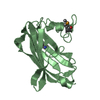
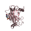
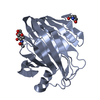
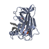
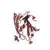
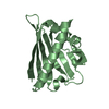
| Entry | Database: PDB / ID: 2agc | ||||||
|---|---|---|---|---|---|---|---|
| Title | Crystal Structure of mouse GM2- activator Protein | ||||||
 Components Components | Ganglioside GM2 activator | ||||||
 Keywords Keywords | LIPID BINDING PROTEIN / constricted lipid binding pocket | ||||||
| Function / homology |  Function and homology information Function and homology informationGlycosphingolipid metabolism /  beta-N-acetylgalactosaminidase activity / positive regulation of hydrolase activity / ganglioside catabolic process / oligosaccharide catabolic process / beta-N-acetylhexosaminidase activity / lipid storage / lipid transporter activity / nervous system process / lipid transport ...Glycosphingolipid metabolism / beta-N-acetylgalactosaminidase activity / positive regulation of hydrolase activity / ganglioside catabolic process / oligosaccharide catabolic process / beta-N-acetylhexosaminidase activity / lipid storage / lipid transporter activity / nervous system process / lipid transport / phospholipase activator activity / neuromuscular process controlling balance / enzyme activator activity / Neutrophil degranulation / cytoplasmic side of plasma membrane / basolateral plasma membrane / lysosome / learning or memory / apical plasma membrane / mitochondrion beta-N-acetylgalactosaminidase activity / positive regulation of hydrolase activity / ganglioside catabolic process / oligosaccharide catabolic process / beta-N-acetylhexosaminidase activity / lipid storage / lipid transporter activity / nervous system process / lipid transport ...Glycosphingolipid metabolism / beta-N-acetylgalactosaminidase activity / positive regulation of hydrolase activity / ganglioside catabolic process / oligosaccharide catabolic process / beta-N-acetylhexosaminidase activity / lipid storage / lipid transporter activity / nervous system process / lipid transport / phospholipase activator activity / neuromuscular process controlling balance / enzyme activator activity / Neutrophil degranulation / cytoplasmic side of plasma membrane / basolateral plasma membrane / lysosome / learning or memory / apical plasma membrane / mitochondrionSimilarity search - Function | ||||||
| Biological species |  Mus musculus (house mouse) Mus musculus (house mouse) | ||||||
| Method | X-RAY DIFFRACTION / MOLECULAR REPLACEMENT / Resolution: 2.5 Å | ||||||
 Authors Authors | Wright, C.S. / Mi, L.Z. / Lee, S. / Rastinejad, F. | ||||||
 Citation Citation | Journal: Biochemistry / Year: 2005 Title: Crystal Structure Analysis of Phosphatidylcholine-GM2-Activator Product Complexes: Evidence for Hydrolase Activity. Authors: Wright, C.S. / Mi, L.Z. / Lee, S. / Rastinejad, F. #1: Journal: J.Mol.Biol. / Year: 2000Title: Crystal Structure of Human GM2- Activator Protein with a Novel beta-cup Topology Authors: Wright, C.S. / Li, S.C. / Rastinejad, F. #2: Journal: J.Mol.Biol. / Year: 2003Title: Structure Analysis of Lipid Complexes of GM2-Activator Protein Authors: Wright, C.S. / Zhao, Q. / Rastinejad, F. #3: Journal: J.Mol.Biol. / Year: 2004Title: Evidence for Lipid Packaging in the Crystal Structure of the GM2-Activator Complex with Platelet Activating Factor Authors: Wright, C.S. / Mi, L.Z. / Rastinejad, F. | ||||||
| History |
|
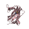
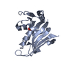
- Structure visualization
| Structure viewer | Molecule: MolmilJmol/JSmol |
|---|
- Downloads & links
-Download
| PDBx/mmCIF format | 2agc.cif.gz | 47.9 KB | Display | PDBx/mmCIF format |
|---|---|---|---|---|
| PDB format | pdb2agc.ent.gz | 32.7 KB | Display | PDB format |
| PDBx/mmJSON format | 2agc.json.gz | Tree view | PDBx/mmJSON format | |
| Others |  Other downloads Other downloads |
-Validation report
| Arichive directory | https://data.pdbj.org/pub/pdb/validation_reports/ag/2agcftp://data.pdbj.org/pub/pdb/validation_reports/ag/2agc | HTTPS FTP |
|---|
-Related structure data
| Related structure data | 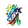 2af9C  2ag2C  2ag4C  2ag9C  1g13S S: Starting model for refinement C: citing same article ( |
|---|---|
| Similar structure data |


-Links
 PDBj
PDBj


- Assembly
| Deposited unit | 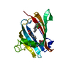
| ||||||||
|---|---|---|---|---|---|---|---|---|---|
| 1 |
| ||||||||
| Unit cell |
|
-Components
| #1: Protein | Mass: 17496.936 Da / Num. of mol.: 1 Source method: isolated from a genetically manipulated source Source: (gene. exp.) Mus musculus (house mouse) / Gene: Gm2a / Organ: brain, kidney, liver / Plasmid: PT7-7 / Species (production host): Escherichia coli / Production host:  Escherichia coli BL21(DE3) (bacteria) / Strain (production host): BL21(DE3) / References: UniProt: Q60648 Escherichia coli BL21(DE3) (bacteria) / Strain (production host): BL21(DE3) / References: UniProt: Q60648 | ||
|---|---|---|---|
| #2: Chemical | ChemComp-MYR / Myristic acid  Mass: 228.371 Da / Num. of mol.: 1 / Source method: obtained synthetically / Formula: C14H28O2 Mass: 228.371 Da / Num. of mol.: 1 / Source method: obtained synthetically / Formula: C14H28O2 | ||
| #3: Chemical | Lauric acid  Mass: 200.318 Da / Num. of mol.: 2 / Source method: obtained synthetically / Formula: C12H24O2 Mass: 200.318 Da / Num. of mol.: 2 / Source method: obtained synthetically / Formula: C12H24O2#4: Water | ChemComp-HOH / | Water Mass: 18.015 Da / Num. of mol.: 66 / Source method: isolated from a natural source / Formula: H2O Mass: 18.015 Da / Num. of mol.: 66 / Source method: isolated from a natural source / Formula: H2O |
-Experimental details
-Experiment
| Experiment | Method: X-RAY DIFFRACTION / Number of used crystals: 1 |
|---|
- Sample preparation
| Crystal | Density Matthews: 2.76 Å3/Da / Density % sol: 47.3 % |
|---|---|
| Crystal grow | Temperature: 298 K / Method: vapor diffusion, hanging drop / pH: 5.6 Details: Peg 4000, acetate buffer , pH 5.6, VAPOR DIFFUSION, HANGING DROP, temperature 298K |
-Data collection
| Diffraction | Mean temperature: 298 K |
|---|---|
| Diffraction source | Source: ROTATING ANODE / Type: RIGAKU / Wavelength: 1.5418 |
| Detector | Type: RIGAKU RAXIS IIC / Detector: IMAGE PLATE / Date: May 2, 1996 / Details: mirrors |
| Radiation | Protocol: SINGLE WAVELENGTH / Monochromatic (M) / Laue (L): M / Scattering type: x-ray |
| Radiation wavelength | Wavelength: 1.5418 Å / Relative weight: 1 |
| Reflection | Resolution: 2.5→50 Å / Num. all: 7535 / Num. obs: 7121 / % possible obs: 94.5 % / Observed criterion σ(F): 1 / Observed criterion σ(I): 1 / Redundancy: 1.8 % / Biso Wilson estimate: 55.1 Å2 / Rmerge(I) obs: 0.06 / Net I/σ(I): 19.3 |
| Reflection shell | Resolution: 2.5→2.59 Å / Redundancy: 1.7 % / Rmerge(I) obs: 0.333 / Mean I/σ(I) obs: 3.1 / Num. unique all: 738 / % possible all: 94.2 |
- Processing
| Software |
| ||||||||||||||||||||
|---|---|---|---|---|---|---|---|---|---|---|---|---|---|---|---|---|---|---|---|---|---|
| Refinement | Method to determine structure: MOLECULAR REPLACEMENT Starting model: 1G13 monomer A Resolution: 2.5→8 Å / Rfactor Rfree error: 0.011 / Data cutoff high absF: 794095.52 / Data cutoff low absF: 0 / Isotropic thermal model: RESTRAINED / Cross valid method: THROUGHOUT / σ(F): 1
| ||||||||||||||||||||
| Solvent computation | Solvent model: FLAT MODEL / Bsol: 80.096 Å2 / ksol: 0.281038 e/Å3 | ||||||||||||||||||||
| Displacement parameters | Biso mean: 52.9 Å2
| ||||||||||||||||||||
| Refine analyze |
| ||||||||||||||||||||
| Refinement step | Cycle: LAST / Resolution: 2.5→8 Å
| ||||||||||||||||||||
| Refine LS restraints |
| ||||||||||||||||||||
| LS refinement shell | Resolution: 2.5→2.66 Å / Rfactor Rfree error: 0.031 / Total num. of bins used: 6
| ||||||||||||||||||||
| Xplor file |
|
