 Movie
Movie Controller

[English] 日本語
 Yorodumi
- PDB-2a9b: Crystal structure of R138Q mutant of recombinant sulfite oxidase ... -
+ Open data

- Basic information
| Entry | Database: PDB / ID: 2a9b | ||||||
|---|---|---|---|---|---|---|---|
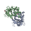
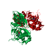
| Title | Crystal structure of R138Q mutant of recombinant sulfite oxidase at resting state | ||||||
 Components Components | Sulfite Oxidase | ||||||
 Keywords Keywords | OXIDOREDUCTASE / sulfite oxidase / molybdopterin / molybdenum / mutant / R160Q / R138Q | ||||||
| Function / homology |  Function and homology informationsulfite oxidase / sulfite oxidase activity / sulfur compound metabolic process / molybdenum ion binding / molybdopterin cofactor binding / mitochondrial intermembrane space / heme binding / mitochondrion Function and homology informationsulfite oxidase / sulfite oxidase activity / sulfur compound metabolic process / molybdenum ion binding / molybdopterin cofactor binding / mitochondrial intermembrane space / heme binding / mitochondrionSimilarity search - Function | ||||||
| Biological species |  Gallus gallus (chicken) Gallus gallus (chicken) | ||||||
| Method | X-RAY DIFFRACTION / SYNCHROTRON / FOURIER SYNTHESIS / Resolution: 2.503 Å | ||||||
 Authors Authors | Karakas, E. / Wilson, H.L. / Graf, T.N. / Xiang, S. / Jaramillo-Busquets, S. / Rajagopalan, K.V. / Kisker, C. | ||||||
 Citation Citation | Journal: J.Biol.Chem. / Year: 2005 Title: Structural insights into sulfite oxidase deficiency Authors: Karakas, E. / Wilson, H.L. / Graf, T.N. / Xiang, S. / Jaramillo-Busquets, S. / Rajagopalan, K.V. / Kisker, C. | ||||||
| History |
|
- Structure visualization
| Structure viewer | Molecule: MolmilJmol/JSmol |
|---|
- Downloads & links
-Download
| PDBx/mmCIF format | 2a9b.cif.gz | 92.5 KB | Display | PDBx/mmCIF format |
|---|---|---|---|---|
| PDB format | pdb2a9b.ent.gz | 68 KB | Display | PDB format |
| PDBx/mmJSON format | 2a9b.json.gz | Tree view | PDBx/mmJSON format | |
| Others |  Other downloads Other downloads |
-Validation report
| Arichive directory | https://data.pdbj.org/pub/pdb/validation_reports/a9/2a9bftp://data.pdbj.org/pub/pdb/validation_reports/a9/2a9b | HTTPS FTP |
|---|
-Related structure data
| Related structure data | 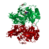 2a99SC 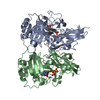 2a9aC  2a9cC 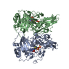 2a9dC S: Starting model for refinement C: citing same article ( |
|---|---|
| Similar structure data |


-Links
 PDBj
PDBj

- Assembly
| Deposited unit | 
| ||||||||||
|---|---|---|---|---|---|---|---|---|---|---|---|
| 1 | 
| ||||||||||
| Unit cell |
| ||||||||||
| Components on special symmetry positions |
| ||||||||||
| Details | The second part of the biological assembly is generated by the two fold axis, 7_544 |
-Components
| #1: Protein | / E.C.1.8.3.1 Mass: 40604.758 Da / Num. of mol.: 1 Fragment: Catalytic core domain and C terminal dimerization domain Mutation: R138Q Source method: isolated from a genetically manipulated source Source: (gene. exp.) Gallus gallus (chicken)Description: The gene is chemically synthesized based on the protein sequence of sulfite oxidase from Gallus gallus Cellular location: Mitochondrial Intermembrane Space MitochondrionGene: SUOX / Organ: Liver / Organelle: Mitochondrion / Plasmid: pTrc99A / Production host:  Escherichia coli (E. coli) / Strain (production host): TP1000 / References: UniProt: P07850, sulfite oxidase Escherichia coli (E. coli) / Strain (production host): TP1000 / References: UniProt: P07850, sulfite oxidase |
|---|---|
| #2: Chemical | ChemComp-CL / Chloride  Mass: 35.453 Da / Num. of mol.: 1 / Source method: obtained synthetically / Formula: Cl Mass: 35.453 Da / Num. of mol.: 1 / Source method: obtained synthetically / Formula: Cl |
| #3: Chemical | ChemComp-MTE /   Mass: 395.352 Da / Num. of mol.: 1 / Source method: obtained synthetically / Formula: C10H14N5O6PS2 Mass: 395.352 Da / Num. of mol.: 1 / Source method: obtained synthetically / Formula: C10H14N5O6PS2 |
| #4: Chemical | ChemComp-MO /   Mass: 95.940 Da / Num. of mol.: 1 / Source method: obtained synthetically / Formula: Mo Mass: 95.940 Da / Num. of mol.: 1 / Source method: obtained synthetically / Formula: Mo |
| #5: Water | ChemComp-HOH / Water Mass: 18.015 Da / Num. of mol.: 215 / Source method: isolated from a natural source / Formula: H2O Mass: 18.015 Da / Num. of mol.: 215 / Source method: isolated from a natural source / Formula: H2O |
-Experimental details
-Experiment
| Experiment | Method: X-RAY DIFFRACTION / Number of used crystals: 1 |
|---|
- Sample preparation
| Crystal | Density Matthews: 3.52 Å3/Da / Density % sol: 64.76 % |
|---|---|
| Crystal grow | Temperature: 295 K / Method: vapor diffusion, hanging drop / pH: 6.5 Details: PEG 4000, magnesium chloride, MES, pH 6.5, VAPOR DIFFUSION, HANGING DROP, temperature 295K |
-Data collection
| Diffraction | Mean temperature: 100 K |
|---|---|
| Diffraction source | Source: SYNCHROTRON / Site: NSLS  / Beamline: X26C / Wavelength: 1.1 Å / Beamline: X26C / Wavelength: 1.1 Å |
| Detector | Type: ADSC QUANTUM 4 / Detector: CCD / Date: Aug 20, 2003 |
| Radiation | Monochromator: Si 111 / Protocol: SINGLE WAVELENGTH / Monochromatic (M) / Laue (L): M / Scattering type: x-ray |
| Radiation wavelength | Wavelength: 1.1 Å / Relative weight: 1 |
| Reflection | Resolution: 2.5→30 Å / Num. obs: 19275 / % possible obs: 100 % / Rmerge(I) obs: 0.089 / Χ2: 1.081 |
| Reflection shell | Resolution: 2.5→2.59 Å / % possible obs: 100 % / Rmerge(I) obs: 0.599 / Num. measured obs: 1936 / Χ2: 1.052 / % possible all: 100 |
- Processing
| Software |
| |||||||||||||||||||||||||||||||||||||||||||||||||||||||||||||||||||||||||||||||||||||||||||||||
|---|---|---|---|---|---|---|---|---|---|---|---|---|---|---|---|---|---|---|---|---|---|---|---|---|---|---|---|---|---|---|---|---|---|---|---|---|---|---|---|---|---|---|---|---|---|---|---|---|---|---|---|---|---|---|---|---|---|---|---|---|---|---|---|---|---|---|---|---|---|---|---|---|---|---|---|---|---|---|---|---|---|---|---|---|---|---|---|---|---|---|---|---|---|---|---|---|
| Refinement | Method to determine structure: FOURIER SYNTHESIS Starting model: PDB ENTRY 2A99 Resolution: 2.503→30 Å / Cor.coef. Fo:Fc: 0.967 / Cor.coef. Fo:Fc free: 0.948 / SU B: 11.74 / SU ML: 0.147 / TLS residual ADP flag: LIKELY RESIDUAL / Cross valid method: THROUGHOUT / σ(F): 0 / ESU R: 0.289 / ESU R Free: 0.212 / Stereochemistry target values: MAXIMUM LIKELIHOOD
| |||||||||||||||||||||||||||||||||||||||||||||||||||||||||||||||||||||||||||||||||||||||||||||||
| Solvent computation | Ion probe radii: 0.8 Å / Shrinkage radii: 0.8 Å / VDW probe radii: 1.2 Å / Solvent model: MASK | |||||||||||||||||||||||||||||||||||||||||||||||||||||||||||||||||||||||||||||||||||||||||||||||
| Displacement parameters | Biso mean: 42.854 Å2
| |||||||||||||||||||||||||||||||||||||||||||||||||||||||||||||||||||||||||||||||||||||||||||||||
| Refinement step | Cycle: LAST / Resolution: 2.503→30 Å
| |||||||||||||||||||||||||||||||||||||||||||||||||||||||||||||||||||||||||||||||||||||||||||||||
| Refine LS restraints |
| |||||||||||||||||||||||||||||||||||||||||||||||||||||||||||||||||||||||||||||||||||||||||||||||
| LS refinement shell | Resolution: 2.503→2.567 Å / Total num. of bins used: 20
| |||||||||||||||||||||||||||||||||||||||||||||||||||||||||||||||||||||||||||||||||||||||||||||||
| Refinement TLS params. | Method: refined / Origin x: 3.208 Å / Origin y: 0.169 Å / Origin z: 18.262 Å
| |||||||||||||||||||||||||||||||||||||||||||||||||||||||||||||||||||||||||||||||||||||||||||||||
| Refinement TLS group | Selection: ALL |
