 Movie
Movie Controller

[English] 日本語
 Yorodumi
- PDB-5hru: Crystal structure of Plasmodium vivax LDH in complex with a DNA a... -
+ Open data

- Basic information
| Entry | Database: PDB / ID: 5hru | |||||||||
|---|---|---|---|---|---|---|---|---|---|---|
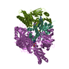
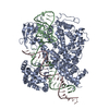
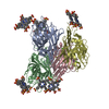
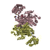
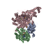
| Title | Crystal structure of Plasmodium vivax LDH in complex with a DNA aptamer called pL1 | |||||||||
 Components Components |
| |||||||||
 Keywords Keywords | OXIDOREDUCTASE/DNA / DNA aptamer / Plasmodium LDH /  malaria / DNA structural element / OXIDOREDUCTASE-DNA complex malaria / DNA structural element / OXIDOREDUCTASE-DNA complex | |||||||||
| Function / homology |  Function and homology information Function and homology informationcarboxylic acid metabolic process / oxidoreductase activity, acting on the CH-OH group of donors, NAD or NADP as acceptor / nucleotide bindingSimilarity search - Function | |||||||||
| Biological species |  Plasmodium vivax (malaria parasite P. vivax) Plasmodium vivax (malaria parasite P. vivax) Phylica emirnensis (plant) Phylica emirnensis (plant) | |||||||||
| Method | X-RAY DIFFRACTION / SYNCHROTRON / MOLECULAR REPLACEMENT / Resolution: 1.71 Å | |||||||||
 Authors Authors | Choi, S.J. / Ban, C. | |||||||||
| Funding support |  Korea, Republic Of, 2items Korea, Republic Of, 2items
| |||||||||
 Citation Citation | Journal: Sci Rep / Year: 2016 Title: Crystal structure of a DNA aptamer bound to PvLDH elucidates novel single-stranded DNA structural elements for folding and recognition Authors: Choi, S.J. / Ban, C. | |||||||||
| History |
|
- Structure visualization
| Structure viewer | Molecule: MolmilJmol/JSmol |
|---|
- Downloads & links
-Download
| PDBx/mmCIF format | 5hru.cif.gz | 286.4 KB | Display | PDBx/mmCIF format |
|---|---|---|---|---|
| PDB format | pdb5hru.ent.gz | 225.6 KB | Display | PDB format |
| PDBx/mmJSON format | 5hru.json.gz | Tree view | PDBx/mmJSON format | |
| Others |  Other downloads Other downloads |
-Validation report
| Arichive directory | https://data.pdbj.org/pub/pdb/validation_reports/hr/5hruftp://data.pdbj.org/pub/pdb/validation_reports/hr/5hru | HTTPS FTP |
|---|
-Related structure data
| Related structure data |  5hs4C  5htoC  2a92S C: citing same article ( S: Starting model for refinement |
|---|---|
| Similar structure data |


-Links
 PDBj
PDBj


- Assembly
| Deposited unit | 
| ||||||||
|---|---|---|---|---|---|---|---|---|---|
| 1 | 
| ||||||||
| Unit cell |
|
-Components
| #1: Protein | Lactate dehydrogenase / Lactate dehydrogenase / Parasite lactate dehydrogenase Mass: 37659.656 Da / Num. of mol.: 2 Source method: isolated from a genetically manipulated source Source: (gene. exp.) Plasmodium vivax (malaria parasite P. vivax)Gene: LDH / Plasmid: pET28a / Production host:  Escherichia coli (E. coli) / References: UniProt: Q4PRK9, L-lactate dehydrogenase Escherichia coli (E. coli) / References: UniProt: Q4PRK9, L-lactate dehydrogenase#2: DNA chain | | Mass: 9944.372 Da / Num. of mol.: 1 / Source method: obtained synthetically / Source: (synth.) Phylica emirnensis (plant)#3: Chemical | ChemComp-MG / |   Mass: 24.305 Da / Num. of mol.: 1 / Source method: obtained synthetically / Formula: Mg Mass: 24.305 Da / Num. of mol.: 1 / Source method: obtained synthetically / Formula: Mg#4: Water | ChemComp-HOH / | Water Mass: 18.015 Da / Num. of mol.: 526 / Source method: isolated from a natural source / Formula: H2O Mass: 18.015 Da / Num. of mol.: 526 / Source method: isolated from a natural source / Formula: H2O |
|---|
-Experimental details
-Experiment
| Experiment | Method: X-RAY DIFFRACTION / Number of used crystals: 1 |
|---|
- Sample preparation
| Crystal | Density Matthews: 2.39 Å3/Da / Density % sol: 43.98 % |
|---|---|
| Crystal grow | Temperature: 293.19 K / Method: vapor diffusion, sitting drop / pH: 7 / Details: PEG-20K, NDSB-195, magnesium chloride |
-Data collection
| Diffraction | Mean temperature: 100 K | |||||||||||||||||||||||||||||||||||||||||||||||||||||||||||||||||||||||||||||||||||||||||||||||||||||||||
|---|---|---|---|---|---|---|---|---|---|---|---|---|---|---|---|---|---|---|---|---|---|---|---|---|---|---|---|---|---|---|---|---|---|---|---|---|---|---|---|---|---|---|---|---|---|---|---|---|---|---|---|---|---|---|---|---|---|---|---|---|---|---|---|---|---|---|---|---|---|---|---|---|---|---|---|---|---|---|---|---|---|---|---|---|---|---|---|---|---|---|---|---|---|---|---|---|---|---|---|---|---|---|---|---|---|---|
| Diffraction source | Source: SYNCHROTRON / Site: PAL/PLS / Beamline: 5C (4A) / Wavelength: 0.9796 Å | |||||||||||||||||||||||||||||||||||||||||||||||||||||||||||||||||||||||||||||||||||||||||||||||||||||||||
| Detector | Type: ADSC QUANTUM 315 / Detector: CCD / Date: Dec 2, 2014 | |||||||||||||||||||||||||||||||||||||||||||||||||||||||||||||||||||||||||||||||||||||||||||||||||||||||||
| Radiation | Protocol: SINGLE WAVELENGTH / Monochromatic (M) / Laue (L): M / Scattering type: x-ray | |||||||||||||||||||||||||||||||||||||||||||||||||||||||||||||||||||||||||||||||||||||||||||||||||||||||||
| Radiation wavelength | Wavelength: 0.9796 Å / Relative weight: 1 | |||||||||||||||||||||||||||||||||||||||||||||||||||||||||||||||||||||||||||||||||||||||||||||||||||||||||
| Reflection | Resolution: 1.71→25 Å / Num. obs: 82531 / % possible obs: 99.8 % / Redundancy: 21.4 % / Rmerge(I) obs: 0.103 / Net I/av σ(I): 80.476 / Net I/σ(I): 15 | |||||||||||||||||||||||||||||||||||||||||||||||||||||||||||||||||||||||||||||||||||||||||||||||||||||||||
| Reflection shell |
|
- Processing
| Software |
| ||||||||||||||||||
|---|---|---|---|---|---|---|---|---|---|---|---|---|---|---|---|---|---|---|---|
| Refinement | Method to determine structure: MOLECULAR REPLACEMENT Starting model: 2A92 Resolution: 1.71→24.96 Å
| ||||||||||||||||||
| Displacement parameters | Biso max: 99.02 Å2 / Biso mean: 24.2256 Å2 / Biso min: 11.36 Å2 | ||||||||||||||||||
| Refinement step | Cycle: LAST / Resolution: 1.71→24.96 Å
| ||||||||||||||||||
| LS refinement shell | Resolution: 1.712→1.773 Å
|
