 Movie
Movie Controller

[English] 日本語
 Yorodumi
- PDB-2a6y: Crystal structure of Emp47p carbohydrate recognition domain (CRD)... -
+ Open data

- Basic information
| Entry | Database: PDB / ID: 2a6y | ||||||
|---|---|---|---|---|---|---|---|
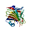
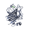
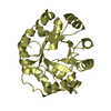
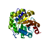
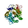
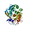
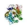
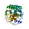
| Title | Crystal structure of Emp47p carbohydrate recognition domain (CRD), tetragonal crystal form | ||||||
 Components Components | Emp47p (form1) | ||||||
 Keywords Keywords | SUGAR BINDING PROTEIN /  BETA SANDWICH / CARBOHYDRATE BINDING PROTEIN / CARGO RECEPTOR / Structural Genomics / NPPSFA / National Project on Protein Structural and Functional Analyses BETA SANDWICH / CARBOHYDRATE BINDING PROTEIN / CARGO RECEPTOR / Structural Genomics / NPPSFA / National Project on Protein Structural and Functional Analyses | ||||||
| Function / homology |  Function and homology information Function and homology informationcarbohydrate derivative binding / fungal-type vacuole membrane / COPII-coated ER to Golgi transport vesicle / D-mannose binding / endoplasmic reticulum-Golgi intermediate compartment / endoplasmic reticulum to Golgi vesicle-mediated transport / Golgi membrane / endoplasmic reticulum membraneSimilarity search - Function | ||||||
| Biological species |  Saccharomyces cerevisiae (brewer's yeast) Saccharomyces cerevisiae (brewer's yeast) | ||||||
| Method | X-RAY DIFFRACTION / SYNCHROTRON / MAD / Resolution: 1.42 Å | ||||||
 Authors Authors | Satoh, T. / Sato, K. / Kanoh, A. / Yamashita, K. / Kato, R. / Nakano, A. / Wakatsuki, S. | ||||||
 Citation Citation | Journal: J.Biol.Chem. / Year: 2006 Title: Structures of the carbohydrate recognition domain of Ca2+-independent cargo receptors Emp46p and Emp47p. Authors: Satoh, T. / Sato, K. / Kanoh, A. / Yamashita, K. / Yamada, Y. / Igarashi, N. / Kato, R. / Nakano, A. / Wakatsuki, S. | ||||||
| History |
|
- Structure visualization
| Structure viewer | Molecule: MolmilJmol/JSmol |
|---|
- Downloads & links
-Download
| PDBx/mmCIF format | 2a6y.cif.gz | 114.2 KB | Display | PDBx/mmCIF format |
|---|---|---|---|---|
| PDB format | pdb2a6y.ent.gz | 93.5 KB | Display | PDB format |
| PDBx/mmJSON format | 2a6y.json.gz | Tree view | PDBx/mmJSON format | |
| Others |  Other downloads Other downloads |
-Validation report
| Arichive directory | https://data.pdbj.org/pub/pdb/validation_reports/a6/2a6yftp://data.pdbj.org/pub/pdb/validation_reports/a6/2a6y | HTTPS FTP |
|---|
-Related structure data
| Related structure data | 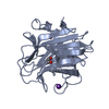 2a6vC  2a6wC  2a6xC 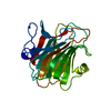 2a6zC  2a70C 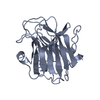 2a71C C: citing same article ( |
|---|---|
| Similar structure data |


-Links
 PDBj
PDBj- Assembly
| Deposited unit | 
| ||||||||||||
|---|---|---|---|---|---|---|---|---|---|---|---|---|---|
| 1 |
| ||||||||||||
| Unit cell |
| ||||||||||||
| Components on special symmetry positions |
|
-Components
| #1: Protein | Mass: 28499.582 Da / Num. of mol.: 1 / Fragment: RESIDUES 1-254 Source method: isolated from a genetically manipulated source Source: (gene. exp.) Saccharomyces cerevisiae (brewer's yeast)Plasmid: pGEX4T-1 / Production host:  Escherichia coli (E. coli) / Strain (production host): BL21(DE3), DL41 / References: GenBank: 854516, UniProt: P43555*PLUS Escherichia coli (E. coli) / Strain (production host): BL21(DE3), DL41 / References: GenBank: 854516, UniProt: P43555*PLUS | ||
|---|---|---|---|
| #2: Chemical | Sulfate  Mass: 96.063 Da / Num. of mol.: 2 / Source method: obtained synthetically / Formula: SO4 Mass: 96.063 Da / Num. of mol.: 2 / Source method: obtained synthetically / Formula: SO4#3: Water | ChemComp-HOH / | Water Mass: 18.015 Da / Num. of mol.: 301 / Source method: isolated from a natural source / Formula: H2O Mass: 18.015 Da / Num. of mol.: 301 / Source method: isolated from a natural source / Formula: H2O |
-Experimental details
-Experiment
| Experiment | Method: X-RAY DIFFRACTION / Number of used crystals: 2 |
|---|
- Sample preparation
| Crystal | Density Matthews: 2.2 Å3/Da / Density % sol: 43.3 % |
|---|---|
| Crystal grow | Temperature: 277 K / Method: vapor diffusion, hanging drop / pH: 6.1 Details: Sodium dihydrogen phosphate, Dipotassium hydrogen phosphate, Lithium sulfate, CAPS, pH 6.1, VAPOR DIFFUSION, HANGING DROP, temperature 277K |
-Data collection
| Diffraction |
| ||||||||||||||||||
|---|---|---|---|---|---|---|---|---|---|---|---|---|---|---|---|---|---|---|---|
| Diffraction source |
| ||||||||||||||||||
| Detector |
| ||||||||||||||||||
| Radiation |
| ||||||||||||||||||
| Radiation wavelength |
| ||||||||||||||||||
| Reflection | Resolution: 1.42→50 Å / Num. all: 48179 / Num. obs: 48001 / % possible obs: 99.6 % / Redundancy: 13.6 % / Biso Wilson estimate: 15.4 Å2 / Rmerge(I) obs: 0.058 / Net I/σ(I): 14 | ||||||||||||||||||
| Reflection shell | Resolution: 1.42→1.47 Å / Redundancy: 13.9 % / Rmerge(I) obs: 0.374 / Mean I/σ(I) obs: 3.6 / Num. unique all: 4735 / % possible all: 99.9 |

- Processing
| Software |
| |||||||||||||||||||||||||||||||||
|---|---|---|---|---|---|---|---|---|---|---|---|---|---|---|---|---|---|---|---|---|---|---|---|---|---|---|---|---|---|---|---|---|---|---|
| Refinement | Method to determine structure: MAD / Resolution: 1.42→10 Å / Num. parameters: 19410 / Num. restraintsaints: 23334 / Cross valid method: THROUGHOUT / σ(F): 0 / Stereochemistry target values: Engh & Huber
| |||||||||||||||||||||||||||||||||
| Refine analyze | Num. disordered residues: 6 / Occupancy sum hydrogen: 0 / Occupancy sum non hydrogen: 2141.5 | |||||||||||||||||||||||||||||||||
| Refinement step | Cycle: LAST / Resolution: 1.42→10 Å
| |||||||||||||||||||||||||||||||||
| Refine LS restraints |
|
