 Movie
Movie Controller

[English] 日本語
 Yorodumi
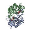
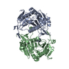
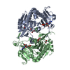
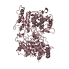
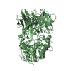
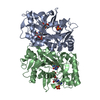
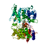
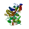
- PDB-1zwj: X-ray structure of galt-like protein from arabidopsis thaliana AT... -
+ Open data

- Basic information
| Entry | Database: PDB / ID: 1zwj | |||||||||
|---|---|---|---|---|---|---|---|---|---|---|
| Title | X-ray structure of galt-like protein from arabidopsis thaliana AT5G18200 | |||||||||
 Components Components | putative galactose-1-phosphate uridyl transferase Galactose-1-phosphate uridylyltransferase Galactose-1-phosphate uridylyltransferase | |||||||||
 Keywords Keywords | STRUCTURAL GENOMICS / UNKNOWN FUNCTION / GALT / AT5G18200 / PROTEIN STRUCTURE INITIATIVE / PSI / CESG / Center for Eukaryotic Structural Genomics | |||||||||
| Function / homology |  Function and homology informationribose-5-phosphate adenylyltransferase activity / positive regulation of cellular response to phosphate starvation / UDP-glucose:hexose-1-phosphate uridylyltransferase activity / galactose catabolic process via UDP-galactose / nucleotidyltransferase activity / ADP binding / glucose metabolic process / Transferases; Transferring phosphorus-containing groups; Nucleotidyltransferases / carbohydrate metabolic process / zinc ion binding Function and homology informationribose-5-phosphate adenylyltransferase activity / positive regulation of cellular response to phosphate starvation / UDP-glucose:hexose-1-phosphate uridylyltransferase activity / galactose catabolic process via UDP-galactose / nucleotidyltransferase activity / ADP binding / glucose metabolic process / Transferases; Transferring phosphorus-containing groups; Nucleotidyltransferases / carbohydrate metabolic process / zinc ion bindingSimilarity search - Function | |||||||||
| Biological species |  Arabidopsis thaliana (thale cress) Arabidopsis thaliana (thale cress) | |||||||||
| Method | X-RAY DIFFRACTION / SYNCHROTRON / MOLECULAR REPLACEMENT, SAD / SAD / Resolution: 2.3 Å | |||||||||
 Authors Authors | Wesenberg, G.E. / Smith, D.W. / Phillips Jr., G.N. / McCoy, J.G. / Johnson, K.A. / Bitto, E. / Bingman, C.A. / Allard, S.T.M. / Center for Eukaryotic Structural Genomics (CESG) | |||||||||
 Citation Citation | Journal: Biochemistry / Year: 2006 Title: Structure and Mechanism of an ADP-Glucose Phosphorylase from Arabidopsis thaliana Authors: MCCOY, J.G. / ARABSHAHI, A. / BITTO, E. / BINGMAN, C.A. / RUZICKA, F.J. / FREY, P.A. / PHILLIPS JR., G.N. | |||||||||
| History |
|
- Structure visualization
| Structure viewer | Molecule: MolmilJmol/JSmol |
|---|
- Downloads & links
-Download
| PDBx/mmCIF format | 1zwj.cif.gz | 138.3 KB | Display | PDBx/mmCIF format |
|---|---|---|---|---|
| PDB format | pdb1zwj.ent.gz | 104.1 KB | Display | PDB format |
| PDBx/mmJSON format | 1zwj.json.gz | Tree view | PDBx/mmJSON format | |
| Others |  Other downloads Other downloads |
-Validation report
| Arichive directory | https://data.pdbj.org/pub/pdb/validation_reports/zw/1zwjftp://data.pdbj.org/pub/pdb/validation_reports/zw/1zwj | HTTPS FTP |
|---|
-Related structure data
| Related structure data |  1z84C  1gupS C: citing same article ( S: Starting model for refinement |
|---|---|
| Similar structure data | |
| Other databases |


-Links
 PDBj
PDBj


- Assembly
| Deposited unit | 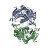
| ||||||||
|---|---|---|---|---|---|---|---|---|---|
| 1 |
| ||||||||
| Unit cell |
| ||||||||
| Details | The biological unit is likely a dimer with one biological unit in the asymmetric unit |
-Components
| #1: Protein | Galactose-1-phosphate uridylyltransferase Mass: 39053.156 Da / Num. of mol.: 2 Source method: isolated from a genetically manipulated source Source: (gene. exp.) Arabidopsis thaliana (thale cress) / Gene: At5g18200 / Plasmid: PVP 13 / Production host:  Escherichia coli (E. coli) / Strain (production host): ROSETTA / References: UniProt: Q9FK51 Escherichia coli (E. coli) / Strain (production host): ROSETTA / References: UniProt: Q9FK51#2: Chemical | ChemComp-ZN /   Mass: 65.409 Da / Num. of mol.: 4 / Source method: obtained synthetically / Formula: Zn Mass: 65.409 Da / Num. of mol.: 4 / Source method: obtained synthetically / Formula: Zn#3: Water | ChemComp-HOH / | Water Mass: 18.015 Da / Num. of mol.: 225 / Source method: isolated from a natural source / Formula: H2O Mass: 18.015 Da / Num. of mol.: 225 / Source method: isolated from a natural source / Formula: H2O |
|---|
-Experimental details
-Experiment
| Experiment | Method: X-RAY DIFFRACTION / Number of used crystals: 3 |
|---|
- Sample preparation
| Crystal | Density Matthews: 2.3 Å3/Da / Density % sol: 47 % |
|---|---|
| Crystal grow | Temperature: 293 K / Method: vapor diffusion, hanging drop / pH: 5.5 Details: 10 MG/ML PROTEIN, 20% PEG 2K, 0.2M SODIUM CHLORIDE, 0.1M MES-ACETATE, pH 5.5, VAPOR DIFFUSION, HANGING DROP, temperature 293K |
-Data collection
| Diffraction |
| ||||||||||||||||||||||||||||||||||||||||||||||||||||||||||||||||||||||||||||||||||||||||||||||||||||||||||||||||||||||||||||||||||||||||||||||||||||||||||||||||||||||||||||||||||||||||||||||||||||||||||||||||||||||||||||||||||||||||||||||||||||||||||||||||||||||||||||||||||||||||||||||||||||||||||||||||||||||||||||||||||
|---|---|---|---|---|---|---|---|---|---|---|---|---|---|---|---|---|---|---|---|---|---|---|---|---|---|---|---|---|---|---|---|---|---|---|---|---|---|---|---|---|---|---|---|---|---|---|---|---|---|---|---|---|---|---|---|---|---|---|---|---|---|---|---|---|---|---|---|---|---|---|---|---|---|---|---|---|---|---|---|---|---|---|---|---|---|---|---|---|---|---|---|---|---|---|---|---|---|---|---|---|---|---|---|---|---|---|---|---|---|---|---|---|---|---|---|---|---|---|---|---|---|---|---|---|---|---|---|---|---|---|---|---|---|---|---|---|---|---|---|---|---|---|---|---|---|---|---|---|---|---|---|---|---|---|---|---|---|---|---|---|---|---|---|---|---|---|---|---|---|---|---|---|---|---|---|---|---|---|---|---|---|---|---|---|---|---|---|---|---|---|---|---|---|---|---|---|---|---|---|---|---|---|---|---|---|---|---|---|---|---|---|---|---|---|---|---|---|---|---|---|---|---|---|---|---|---|---|---|---|---|---|---|---|---|---|---|---|---|---|---|---|---|---|---|---|---|---|---|---|---|---|---|---|---|---|---|---|---|---|---|---|---|---|---|---|---|---|---|---|---|---|---|---|---|---|---|---|---|---|---|---|---|---|---|---|---|---|---|---|---|---|---|---|---|---|---|---|---|---|---|---|---|---|---|---|---|---|---|---|---|---|---|---|---|---|---|---|---|---|---|---|---|---|
| Diffraction source |
| ||||||||||||||||||||||||||||||||||||||||||||||||||||||||||||||||||||||||||||||||||||||||||||||||||||||||||||||||||||||||||||||||||||||||||||||||||||||||||||||||||||||||||||||||||||||||||||||||||||||||||||||||||||||||||||||||||||||||||||||||||||||||||||||||||||||||||||||||||||||||||||||||||||||||||||||||||||||||||||||||||
| Detector |
| ||||||||||||||||||||||||||||||||||||||||||||||||||||||||||||||||||||||||||||||||||||||||||||||||||||||||||||||||||||||||||||||||||||||||||||||||||||||||||||||||||||||||||||||||||||||||||||||||||||||||||||||||||||||||||||||||||||||||||||||||||||||||||||||||||||||||||||||||||||||||||||||||||||||||||||||||||||||||||||||||||
| Radiation |
| ||||||||||||||||||||||||||||||||||||||||||||||||||||||||||||||||||||||||||||||||||||||||||||||||||||||||||||||||||||||||||||||||||||||||||||||||||||||||||||||||||||||||||||||||||||||||||||||||||||||||||||||||||||||||||||||||||||||||||||||||||||||||||||||||||||||||||||||||||||||||||||||||||||||||||||||||||||||||||||||||||
| Radiation wavelength |
| ||||||||||||||||||||||||||||||||||||||||||||||||||||||||||||||||||||||||||||||||||||||||||||||||||||||||||||||||||||||||||||||||||||||||||||||||||||||||||||||||||||||||||||||||||||||||||||||||||||||||||||||||||||||||||||||||||||||||||||||||||||||||||||||||||||||||||||||||||||||||||||||||||||||||||||||||||||||||||||||||||
| Reflection | D res low: 50 Å
| ||||||||||||||||||||||||||||||||||||||||||||||||||||||||||||||||||||||||||||||||||||||||||||||||||||||||||||||||||||||||||||||||||||||||||||||||||||||||||||||||||||||||||||||||||||||||||||||||||||||||||||||||||||||||||||||||||||||||||||||||||||||||||||||||||||||||||||||||||||||||||||||||||||||||||||||||||||||||||||||||||
| Diffraction reflection shell |
| ||||||||||||||||||||||||||||||||||||||||||||||||||||||||||||||||||||||||||||||||||||||||||||||||||||||||||||||||||||||||||||||||||||||||||||||||||||||||||||||||||||||||||||||||||||||||||||||||||||||||||||||||||||||||||||||||||||||||||||||||||||||||||||||||||||||||||||||||||||||||||||||||||||||||||||||||||||||||||||||||||
| Reflection | Resolution: 2.3→50 Å / Num. obs: 27575 / % possible obs: 95.6 % / Redundancy: 6.4 % / Rmerge(I) obs: 0.044 / Χ2: 0.858 / Net I/σ(I): 24.03 | ||||||||||||||||||||||||||||||||||||||||||||||||||||||||||||||||||||||||||||||||||||||||||||||||||||||||||||||||||||||||||||||||||||||||||||||||||||||||||||||||||||||||||||||||||||||||||||||||||||||||||||||||||||||||||||||||||||||||||||||||||||||||||||||||||||||||||||||||||||||||||||||||||||||||||||||||||||||||||||||||||
| Reflection shell | Diffraction-ID: 1,2,3
|

-Phasing
| Phasing | Method: SAD | ||||||||||||||||||||||||||||||||||||||||||||||||||||||||||||||||||||||||||||||||||||||||||||||||||||||||||||||||||||||||||||||
|---|---|---|---|---|---|---|---|---|---|---|---|---|---|---|---|---|---|---|---|---|---|---|---|---|---|---|---|---|---|---|---|---|---|---|---|---|---|---|---|---|---|---|---|---|---|---|---|---|---|---|---|---|---|---|---|---|---|---|---|---|---|---|---|---|---|---|---|---|---|---|---|---|---|---|---|---|---|---|---|---|---|---|---|---|---|---|---|---|---|---|---|---|---|---|---|---|---|---|---|---|---|---|---|---|---|---|---|---|---|---|---|---|---|---|---|---|---|---|---|---|---|---|---|---|---|---|---|
| Phasing MAD | D res high: 3.49 Å / D res low: 40.19 Å / FOM : 0.321 / FOM acentric: 0.353 / FOM centric: 0 / Reflection: 15287 | ||||||||||||||||||||||||||||||||||||||||||||||||||||||||||||||||||||||||||||||||||||||||||||||||||||||||||||||||||||||||||||||
| Phasing MAD set | R cullis: 0.673 / R cullis acentric: 0.64 / R cullis centric: 10 / R kraut: 0.032 / R kraut acentric: 0.032 / R kraut centric: 0.035 / Highest resolution: 3.49 Å / Lowest resolution: 40.19 Å / FOM : 0.319 / FOM acentric: 0.351 / FOM centric: 0 / Loc: 5.185 / Loc acentric: 5.18 / Loc centric: 5.282 / Power: 1.634 kW / Power acentric: 1.604 / Power centric: 1.903 | ||||||||||||||||||||||||||||||||||||||||||||||||||||||||||||||||||||||||||||||||||||||||||||||||||||||||||||||||||||||||||||||
| Phasing MAD set | Wavelength: 0.97908 Å / Atom type: SE / F double prime refined: 0.68 / F prime refined: -10 | ||||||||||||||||||||||||||||||||||||||||||||||||||||||||||||||||||||||||||||||||||||||||||||||||||||||||||||||||||||||||||||||
| Phasing MAD set shell | ID: 1 / R cullis centric: 10 / FOM centric: 0
| ||||||||||||||||||||||||||||||||||||||||||||||||||||||||||||||||||||||||||||||||||||||||||||||||||||||||||||||||||||||||||||||
| Phasing MAD set site |
| ||||||||||||||||||||||||||||||||||||||||||||||||||||||||||||||||||||||||||||||||||||||||||||||||||||||||||||||||||||||||||||||
| Phasing MAD shell |
|
- Processing
| Software |
| ||||||||||||||||||||||||||||||||||||||||||||||||||||||||||||||||||||||||||||||||||||||||||
|---|---|---|---|---|---|---|---|---|---|---|---|---|---|---|---|---|---|---|---|---|---|---|---|---|---|---|---|---|---|---|---|---|---|---|---|---|---|---|---|---|---|---|---|---|---|---|---|---|---|---|---|---|---|---|---|---|---|---|---|---|---|---|---|---|---|---|---|---|---|---|---|---|---|---|---|---|---|---|---|---|---|---|---|---|---|---|---|---|---|---|---|
| Refinement | Method to determine structure: MOLECULAR REPLACEMENT, SAD Starting model: PDB ENTRY 1GUP Resolution: 2.3→27.59 Å / Rfactor Rfree error: 0.005 / Occupancy max: 1 / Occupancy min: 1 / Cross valid method: THROUGHOUT
| ||||||||||||||||||||||||||||||||||||||||||||||||||||||||||||||||||||||||||||||||||||||||||
| Solvent computation | Solvent model: CNS bulk solvent model used / Bsol: 37.3753 Å2 / ksol: 0.327623 e/Å3 | ||||||||||||||||||||||||||||||||||||||||||||||||||||||||||||||||||||||||||||||||||||||||||
| Displacement parameters | Biso max: 100.34 Å2 / Biso mean: 47.15 Å2 / Biso min: 19.99 Å2
| ||||||||||||||||||||||||||||||||||||||||||||||||||||||||||||||||||||||||||||||||||||||||||
| Refine analyze |
| ||||||||||||||||||||||||||||||||||||||||||||||||||||||||||||||||||||||||||||||||||||||||||
| Refinement step | Cycle: LAST / Resolution: 2.3→27.59 Å
| ||||||||||||||||||||||||||||||||||||||||||||||||||||||||||||||||||||||||||||||||||||||||||
| Refine LS restraints |
| ||||||||||||||||||||||||||||||||||||||||||||||||||||||||||||||||||||||||||||||||||||||||||
| LS refinement shell | Refine-ID: X-RAY DIFFRACTION / Total num. of bins used: 8
| ||||||||||||||||||||||||||||||||||||||||||||||||||||||||||||||||||||||||||||||||||||||||||
| Xplor file |
|
