 Movie
Movie Controller

[English] 日本語
 Yorodumi
- PDB-1zvy: Crystal structure of the VHH D3-L11 in complex with hen egg white... -
+ Open data

- Basic information
| Entry | Database: PDB / ID: 1zvy | ||||||
|---|---|---|---|---|---|---|---|
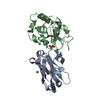
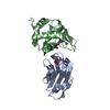
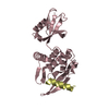
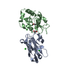
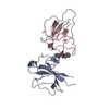
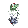
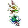
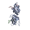
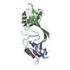
| Title | Crystal structure of the VHH D3-L11 in complex with hen egg white lysozyme | ||||||
 Components Components |
| ||||||
 Keywords Keywords | HYDROLASE/IMMUNE SYSTEM /  beta sandwich / immunoglobulin fold / protein protein heterocomplex / alpha-beta orthogonal bundle / HYDROLASE-IMMUNE SYSTEM COMPLEX beta sandwich / immunoglobulin fold / protein protein heterocomplex / alpha-beta orthogonal bundle / HYDROLASE-IMMUNE SYSTEM COMPLEX | ||||||
| Function / homology |  Function and homology information Function and homology informationLactose synthesis / Antimicrobial peptides / Neutrophil degranulation / beta-N-acetylglucosaminidase activity / cell wall macromolecule catabolic process / lysozyme / lysozyme activity / killing of cells of another organism / defense response to Gram-negative bacterium / defense response to Gram-positive bacterium ...Lactose synthesis / Antimicrobial peptides / Neutrophil degranulation / beta-N-acetylglucosaminidase activity / cell wall macromolecule catabolic process / lysozyme / lysozyme activity / killing of cells of another organism / defense response to Gram-negative bacterium / defense response to Gram-positive bacterium / defense response to bacterium / endoplasmic reticulum / extracellular space / identical protein binding / cytoplasmSimilarity search - Function | ||||||
| Biological species |  Camelus dromedarius (Arabian camel) Camelus dromedarius (Arabian camel) Gallus gallus (chicken) Gallus gallus (chicken) | ||||||
| Method | X-RAY DIFFRACTION / SYNCHROTRON / MOLECULAR REPLACEMENT / Resolution: 1.63 Å | ||||||
 Authors Authors | De Genst, E. / Silence, K. / Decanniere, K. / Conrath, K. / Loris, R. / Kinne, J. / Muyldermans, S. / Wyns, L. | ||||||
 Citation Citation | Journal: Proc.Natl.Acad.Sci.Usa / Year: 2006 Title: Molecular basis for the preferential cleft recognition by dromedary heavy-chain antibodies. Authors: De Genst, E. / Silence, K. / Decanniere, K. / Conrath, K. / Loris, R. / Kinne, J. / Muyldermans, S. / Wyns, L. | ||||||
| History |
| ||||||
| Remark 999 | SEQUENCE The sequence of a camelid heavy-chain antibody has not been deposited into any sequence database. |
- Structure visualization
| Structure viewer | Molecule: MolmilJmol/JSmol |
|---|
- Downloads & links
-Download
| PDBx/mmCIF format | 1zvy.cif.gz | 67.3 KB | Display | PDBx/mmCIF format |
|---|---|---|---|---|
| PDB format | pdb1zvy.ent.gz | 47.9 KB | Display | PDB format |
| PDBx/mmJSON format | 1zvy.json.gz | Tree view | PDBx/mmJSON format | |
| Others |  Other downloads Other downloads |
-Validation report
| Arichive directory | https://data.pdbj.org/pub/pdb/validation_reports/zv/1zvyftp://data.pdbj.org/pub/pdb/validation_reports/zv/1zvy | HTTPS FTP |
|---|
-Related structure data
| Related structure data |  1zv5C 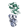 1zvhC 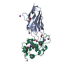 1jttS C: citing same article ( S: Starting model for refinement |
|---|---|
| Similar structure data |

-Links
 PDBj
PDBj


- Assembly
| Deposited unit | 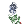
| ||||||||
|---|---|---|---|---|---|---|---|---|---|
| 1 |
| ||||||||
| Unit cell |
|
-Components
| #1: Antibody | Mass: 14994.540 Da / Num. of mol.: 1 / Fragment: D3-L11 Source method: isolated from a genetically manipulated source Source: (gene. exp.) Camelus dromedarius (Arabian camel) / Plasmid: pHEN06 / Production host:  Escherichia coli (E. coli) / Strain (production host): WK6Su- Escherichia coli (E. coli) / Strain (production host): WK6Su- |
|---|---|
| #2: Protein | Mass: 14331.160 Da / Num. of mol.: 1 / Source method: isolated from a natural source / Source: (natural) Gallus gallus (chicken) / References: UniProt: P00698, lysozyme |
| #3: Chemical | ChemComp-PO4 / Phosphate  Mass: 94.971 Da / Num. of mol.: 1 / Source method: obtained synthetically / Formula: PO4 Mass: 94.971 Da / Num. of mol.: 1 / Source method: obtained synthetically / Formula: PO4 |
| #4: Chemical | ChemComp-GOL / Glycerol  Mass: 92.094 Da / Num. of mol.: 1 / Source method: obtained synthetically / Formula: C3H8O3 Mass: 92.094 Da / Num. of mol.: 1 / Source method: obtained synthetically / Formula: C3H8O3 |
| #5: Water | ChemComp-HOH / Water Mass: 18.015 Da / Num. of mol.: 218 / Source method: isolated from a natural source / Formula: H2O Mass: 18.015 Da / Num. of mol.: 218 / Source method: isolated from a natural source / Formula: H2O |
-Experimental details
-Experiment
| Experiment | Method: X-RAY DIFFRACTION / Number of used crystals: 1 |
|---|
- Sample preparation
| Crystal | Density Matthews: 1.9 Å3/Da / Density % sol: 34.9 % |
|---|---|
| Crystal grow | Temperature: 293 K / Method: vapor diffusion, hanging drop / pH: 6.5 Details: magnesium acetate, sodium cacodylate, PEG 8000, glycerol, pH 6.5, VAPOR DIFFUSION, HANGING DROP, temperature 293K |
-Data collection
| Diffraction | Mean temperature: 100 K |
|---|---|
| Diffraction source | Source: SYNCHROTRON / Site: EMBL/DESY, HAMBURG  / Beamline: BW7B / Wavelength: 0.8453 Å / Beamline: BW7B / Wavelength: 0.8453 Å |
| Detector | Type: MARRESEARCH / Detector: IMAGE PLATE / Date: Jan 20, 2002 |
| Radiation | Monochromator: SAGITALLY FOCUSED Si(111) / Protocol: SINGLE WAVELENGTH / Monochromatic (M) / Laue (L): M / Scattering type: x-ray |
| Radiation wavelength | Wavelength: 0.8453 Å / Relative weight: 1 |
| Reflection | Resolution: 1.6→30 Å / Num. all: 29964 / Num. obs: 29801 / % possible obs: 99.5 % / Observed criterion σ(F): 0 / Observed criterion σ(I): 0 / Rmerge(I) obs: 0.087 / Χ2: 1.308 |
| Reflection shell | Resolution: 1.6→1.66 Å / % possible obs: 98.8 % / Rmerge(I) obs: 0.354 / Num. measured obs: 2940 / Χ2: 0.954 / % possible all: 98.8 |
- Processing
| Software |
| ||||||||||||||||||||||||||||||||||||
|---|---|---|---|---|---|---|---|---|---|---|---|---|---|---|---|---|---|---|---|---|---|---|---|---|---|---|---|---|---|---|---|---|---|---|---|---|---|
| Refinement | Method to determine structure: MOLECULAR REPLACEMENT Starting model: pdb entry 1JTT Resolution: 1.63→17.74 Å / Rfactor Rfree error: 0.006 / Data cutoff high absF: 1032625.25 / Data cutoff low absF: 0 / Isotropic thermal model: RESTRAINED / Cross valid method: THROUGHOUT / σ(F): 0 / Stereochemistry target values: Engh & Huber / Details: BULK SOLVENT MODEL USED
| ||||||||||||||||||||||||||||||||||||
| Solvent computation | Solvent model: FLAT MODEL / Bsol: 61.781 Å2 / ksol: 0.443 e/Å3 | ||||||||||||||||||||||||||||||||||||
| Displacement parameters | Biso mean: 19.3 Å2
| ||||||||||||||||||||||||||||||||||||
| Refine analyze |
| ||||||||||||||||||||||||||||||||||||
| Refinement step | Cycle: LAST / Resolution: 1.63→17.74 Å
| ||||||||||||||||||||||||||||||||||||
| Refine LS restraints |
| ||||||||||||||||||||||||||||||||||||
| Refine LS restraints NCS | NCS model details: RESTRAINTS | ||||||||||||||||||||||||||||||||||||
| LS refinement shell | Resolution: 1.63→1.7 Å / Rfactor Rfree error: 0.024 / Total num. of bins used: 6
| ||||||||||||||||||||||||||||||||||||
| Xplor file |
|
