 Movie
Movie Controller

[English] 日本語
 Yorodumi
- PDB-1q3p: Crystal structure of the Shank PDZ-ligand complex reveals a class... -
+ Open data

- Basic information
| Entry | Database: PDB / ID: 1q3p | ||||||
|---|---|---|---|---|---|---|---|
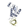
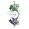
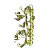
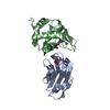
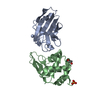
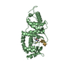
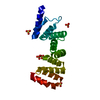
| Title | Crystal structure of the Shank PDZ-ligand complex reveals a class I PDZ interaction and a novel PDZ-PDZ dimerization | ||||||
 Components Components |
| ||||||
 Keywords Keywords | PEPTIDE BINDING PROTEIN / shank / PDZ /  GKAP GKAP | ||||||
| Function / homology |  Function and homology informationsomatostatin receptor binding / determination of affect / synaptic receptor adaptor activity / olfactory behavior / synapse maturation / Neurexins and neuroligins / negative regulation of actin filament bundle assembly / structural constituent of postsynaptic density / righting reflex / protein localization to synapse ...somatostatin receptor binding / determination of affect / synaptic receptor adaptor activity / olfactory behavior / synapse maturation / Neurexins and neuroligins / negative regulation of actin filament bundle assembly / structural constituent of postsynaptic density / righting reflex / protein localization to synapse / vocalization behavior / habituation / regulation of AMPA receptor activity / ankyrin repeat binding / dendritic spine morphogenesis / adult behavior / positive regulation of dendritic spine development / social behavior / associative learning / positive regulation of excitatory postsynaptic potential / neuromuscular process controlling balance / excitatory synapse / long-term memory / ionotropic glutamate receptor binding / G protein-coupled receptor binding / modulation of chemical synaptic transmission / Schaffer collateral - CA1 synapse / SH3 domain binding / signaling receptor complex adaptor activity / scaffold protein binding / postsynaptic membrane / protein-containing complex assembly / dendritic spine / postsynaptic density / neuron projection / dendrite / synapse / glutamatergic synapse / protein-containing complex binding / membrane / identical protein binding / plasma membrane / cytosol Function and homology informationsomatostatin receptor binding / determination of affect / synaptic receptor adaptor activity / olfactory behavior / synapse maturation / Neurexins and neuroligins / negative regulation of actin filament bundle assembly / structural constituent of postsynaptic density / righting reflex / protein localization to synapse ...somatostatin receptor binding / determination of affect / synaptic receptor adaptor activity / olfactory behavior / synapse maturation / Neurexins and neuroligins / negative regulation of actin filament bundle assembly / structural constituent of postsynaptic density / righting reflex / protein localization to synapse / vocalization behavior / habituation / regulation of AMPA receptor activity / ankyrin repeat binding / dendritic spine morphogenesis / adult behavior / positive regulation of dendritic spine development / social behavior / associative learning / positive regulation of excitatory postsynaptic potential / neuromuscular process controlling balance / excitatory synapse / long-term memory / ionotropic glutamate receptor binding / G protein-coupled receptor binding / modulation of chemical synaptic transmission / Schaffer collateral - CA1 synapse / SH3 domain binding / signaling receptor complex adaptor activity / scaffold protein binding / postsynaptic membrane / protein-containing complex assembly / dendritic spine / postsynaptic density / neuron projection / dendrite / synapse / glutamatergic synapse / protein-containing complex binding / membrane / identical protein binding / plasma membrane / cytosolSimilarity search - Function | ||||||
| Biological species |  Rattus norvegicus (Norway rat) Rattus norvegicus (Norway rat) | ||||||
| Method | X-RAY DIFFRACTION / SYNCHROTRON / MOLECULAR REPLACEMENT / Resolution: 2.25 Å | ||||||
 Authors Authors | Im, Y.J. / Lee, J.H. / Park, S.H. / Park, S.J. / Rho, S.-H. / Kang, G.B. / Kim, E. / Eom, S.H. | ||||||
 Citation Citation | Journal: J.Biol.Chem. / Year: 2003 Title: Crystal structure of the Shank PDZ-ligand complex reveals a class I PDZ interaction and a novel PDZ-PDZ dimerization Authors: Im, Y.J. / Lee, J.H. / Park, S.H. / Park, S.J. / Rho, S.-H. / Kang, G.B. / Kim, E. / Eom, S.H. | ||||||
| History |
|
- Structure visualization
| Structure viewer | Molecule: MolmilJmol/JSmol |
|---|
- Downloads & links
-Download
| PDBx/mmCIF format | 1q3p.cif.gz | 54.4 KB | Display | PDBx/mmCIF format |
|---|---|---|---|---|
| PDB format | pdb1q3p.ent.gz | 39.3 KB | Display | PDB format |
| PDBx/mmJSON format | 1q3p.json.gz | Tree view | PDBx/mmJSON format | |
| Others |  Other downloads Other downloads |
-Validation report
| Arichive directory | https://data.pdbj.org/pub/pdb/validation_reports/q3/1q3pftp://data.pdbj.org/pub/pdb/validation_reports/q3/1q3p | HTTPS FTP |
|---|
-Related structure data
| Related structure data |  1q3oSC S: Starting model for refinement C: citing same article ( |
|---|---|
| Similar structure data |
-Links
 PDBj
PDBj- Assembly
| Deposited unit | 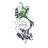
| ||||||||
|---|---|---|---|---|---|---|---|---|---|
| 1 |
| ||||||||
| 2 | 
| ||||||||
| Unit cell |
|
-Components
| #1: Protein | Mass: 12073.959 Da / Num. of mol.: 2 / Fragment: PDZ domain Source method: isolated from a genetically manipulated source Source: (gene. exp.) Rattus norvegicus (Norway rat) / Gene: shank1 / Plasmid: pGEX4T1 / Species (production host): Escherichia coli / Production host:  Escherichia coli BL21(DE3) (bacteria) / Strain (production host): BL21(DE3) / References: UniProt: Q9WV48 Escherichia coli BL21(DE3) (bacteria) / Strain (production host): BL21(DE3) / References: UniProt: Q9WV48#2: Protein/peptide | Mass: 717.791 Da / Num. of mol.: 2 / Source method: obtained synthetically Details: EAQTRL sequence is the C-terminal hexapeptide of GKAP protein in rattus norvegicus References: GenBank: 19923689 #3: Water | ChemComp-HOH / | Water Mass: 18.015 Da / Num. of mol.: 44 / Source method: isolated from a natural source / Formula: H2O Mass: 18.015 Da / Num. of mol.: 44 / Source method: isolated from a natural source / Formula: H2O |
|---|
-Experimental details
-Experiment
| Experiment | Method: X-RAY DIFFRACTION / Number of used crystals: 1 |
|---|
- Sample preparation
| Crystal | Density Matthews: 3.04 Å3/Da / Density % sol: 59.25 % | |||||||||||||||||||||||||||||||||||||||||||||||||
|---|---|---|---|---|---|---|---|---|---|---|---|---|---|---|---|---|---|---|---|---|---|---|---|---|---|---|---|---|---|---|---|---|---|---|---|---|---|---|---|---|---|---|---|---|---|---|---|---|---|---|
| Crystal grow | Temperature: 294 K / Method: vapor diffusion, hanging drop / pH: 6.5 Details: PEG 6000, KCl, pH 6.5, VAPOR DIFFUSION, HANGING DROP, temperature 294K | |||||||||||||||||||||||||||||||||||||||||||||||||
| Crystal grow | *PLUS pH: 7.4 / Method: vapor diffusion, hanging drop / Details: Park, S.H., (2002) Acta Cryst., D58, 1353. | |||||||||||||||||||||||||||||||||||||||||||||||||
| Components of the solutions | *PLUS
|
-Data collection
| Diffraction | Mean temperature: 100 K |
|---|---|
| Diffraction source | Source: SYNCHROTRON / Site: PAL/PLS  / Beamline: 6B / Wavelength: 1 Å / Beamline: 6B / Wavelength: 1 Å |
| Detector | Type: MARRESEARCH / Detector: IMAGE PLATE / Date: Apr 26, 2002 / Details: mirrors |
| Radiation | Monochromator: Si 111 CHANNEL / Protocol: SINGLE WAVELENGTH / Monochromatic (M) / Laue (L): M / Scattering type: x-ray |
| Radiation wavelength | Wavelength: 1 Å / Relative weight: 1 |
| Reflection | Resolution: 2.25→30 Å / Num. all: 64296 / Num. obs: 61853 / % possible obs: 96.2 % / Observed criterion σ(F): 2 / Observed criterion σ(I): 4 / Redundancy: 4.3 % / Biso Wilson estimate: 30.3 Å2 / Rmerge(I) obs: 0.041 / Rsym value: 0.041 / Net I/σ(I): 50.9 |
| Reflection shell | Resolution: 2.25→2.29 Å / Rmerge(I) obs: 0.258 / Mean I/σ(I) obs: 6.6 / Rsym value: 0.258 / % possible all: 96.5 |
| Reflection shell | *PLUS % possible obs: 95.6 % |
- Processing
| Software |
| ||||||||||||||||||||||||||||||||||||||||||||||||||||||||||||||||||||||||||||||||
|---|---|---|---|---|---|---|---|---|---|---|---|---|---|---|---|---|---|---|---|---|---|---|---|---|---|---|---|---|---|---|---|---|---|---|---|---|---|---|---|---|---|---|---|---|---|---|---|---|---|---|---|---|---|---|---|---|---|---|---|---|---|---|---|---|---|---|---|---|---|---|---|---|---|---|---|---|---|---|---|---|---|
| Refinement | Method to determine structure: MOLECULAR REPLACEMENT Starting model: PDB entry 1Q3O Resolution: 2.25→29.84 Å / Rfactor Rfree error: 0.01 / Data cutoff high absF: 840523.46 / Data cutoff low absF: 0 / Isotropic thermal model: RESTRAINED / Cross valid method: THROUGHOUT / σ(F): 0 / σ(I): 0 / Stereochemistry target values: Engh & Huber
| ||||||||||||||||||||||||||||||||||||||||||||||||||||||||||||||||||||||||||||||||
| Solvent computation | Solvent model: FLAT MODEL / Bsol: 52.5189 Å2 / ksol: 0.359095 e/Å3 | ||||||||||||||||||||||||||||||||||||||||||||||||||||||||||||||||||||||||||||||||
| Displacement parameters | Biso mean: 44.1 Å2
| ||||||||||||||||||||||||||||||||||||||||||||||||||||||||||||||||||||||||||||||||
| Refine analyze |
| ||||||||||||||||||||||||||||||||||||||||||||||||||||||||||||||||||||||||||||||||
| Refinement step | Cycle: LAST / Resolution: 2.25→29.84 Å
| ||||||||||||||||||||||||||||||||||||||||||||||||||||||||||||||||||||||||||||||||
| Refine LS restraints |
| ||||||||||||||||||||||||||||||||||||||||||||||||||||||||||||||||||||||||||||||||
| LS refinement shell | Resolution: 2.25→2.39 Å / Rfactor Rfree error: 0.028 / Total num. of bins used: 6
| ||||||||||||||||||||||||||||||||||||||||||||||||||||||||||||||||||||||||||||||||
| Xplor file |
| ||||||||||||||||||||||||||||||||||||||||||||||||||||||||||||||||||||||||||||||||
| Refinement | *PLUS Lowest resolution: 30 Å / % reflection Rfree: 5 % / Rfactor Rfree: 0.28 | ||||||||||||||||||||||||||||||||||||||||||||||||||||||||||||||||||||||||||||||||
| Solvent computation | *PLUS | ||||||||||||||||||||||||||||||||||||||||||||||||||||||||||||||||||||||||||||||||
| Displacement parameters | *PLUS | ||||||||||||||||||||||||||||||||||||||||||||||||||||||||||||||||||||||||||||||||
| Refine LS restraints | *PLUS
|
