 Movie
Movie Controller

[English] 日本語
 Yorodumi
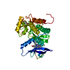
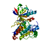
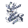
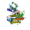
- PDB-1zgy: Structural and Biochemical Basis for Selective Repression of the ... -
+ Open data

- Basic information
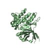
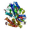
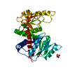
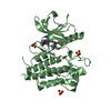
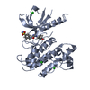
| Entry | Database: PDB / ID: 1zgy | ||||||
|---|---|---|---|---|---|---|---|
| Title | Structural and Biochemical Basis for Selective Repression of the Orphan Nuclear Receptor LRH-1 by SHP | ||||||
 Components Components |
| ||||||
 Keywords Keywords |  TRANSCRIPTION / protein-peptide complex TRANSCRIPTION / protein-peptide complex | ||||||
| Function / homology |  Function and homology information Function and homology informationNuclear Receptor transcription pathway / nuclear retinoic acid receptor binding / bile acid and bile salt transport / prostaglandin receptor activity / regulation of cholesterol transporter activity / negative regulation of connective tissue replacement involved in inflammatory response wound healing / negative regulation of receptor signaling pathway via STAT / transcription regulator inhibitor activity / peroxisome proliferator activated receptor binding / MECP2 regulates transcription factors ...Nuclear Receptor transcription pathway / nuclear retinoic acid receptor binding / bile acid and bile salt transport / prostaglandin receptor activity / regulation of cholesterol transporter activity / negative regulation of connective tissue replacement involved in inflammatory response wound healing / negative regulation of receptor signaling pathway via STAT / transcription regulator inhibitor activity / peroxisome proliferator activated receptor binding / MECP2 regulates transcription factors / negative regulation of extracellular matrix assembly / negative regulation of vascular endothelial cell proliferation / negative regulation of cellular response to transforming growth factor beta stimulus / negative regulation of cardiac muscle hypertrophy in response to stress / arachidonic acid binding / nuclear thyroid hormone receptor binding / positive regulation of low-density lipoprotein receptor activity / positive regulation of adiponectin secretion / lipoprotein transport / negative regulation of sequestering of triglyceride / macrophage derived foam cell differentiation / positive regulation of vascular associated smooth muscle cell apoptotic process / DNA binding domain binding / STAT family protein binding / positive regulation of fatty acid metabolic process / response to lipid / negative regulation of SMAD protein signal transduction / LBD domain binding / negative regulation of type II interferon-mediated signaling pathway / negative regulation of cholesterol storage / E-box binding / alpha-actinin binding / lipid homeostasis / negative regulation of vascular associated smooth muscle cell proliferation / R-SMAD binding / monocyte differentiation / negative regulation of macrophage derived foam cell differentiation / cellular response to low-density lipoprotein particle stimulus / negative regulation of lipid storage / negative regulation of blood vessel endothelial cell migration / negative regulation of BMP signaling pathway / white fat cell differentiation / negative regulation of mitochondrial fission / positive regulation of cholesterol efflux / retinoic acid receptor signaling pathway / positive regulation of fat cell differentiation / negative regulation of osteoblast differentiation / cell fate commitment / positive regulation of DNA binding / animal organ regeneration / BMP signaling pathway / long-chain fatty acid transport / response to glucose / nuclear retinoid X receptor binding / negative regulation of signaling receptor activity / regulation of cellular response to insulin stimulus / cell maturation / Notch signaling pathway / epithelial cell differentiation / positive regulation of adipose tissue development / peroxisome proliferator activated receptor signaling pathway / hormone-mediated signaling pathway / negative regulation of angiogenesis / response to nutrient / negative regulation of miRNA transcription / negative regulation of MAP kinase activity / fatty acid metabolic process / Regulation of PTEN gene transcription / transcription coregulator binding / placenta development / negative regulation of smooth muscle cell proliferation / peptide binding / negative regulation of transforming growth factor beta receptor signaling pathway / circadian regulation of gene expression / SUMOylation of intracellular receptors / mRNA transcription by RNA polymerase II / regulation of circadian rhythm / positive regulation of insulin secretion / lipid metabolic process / negative regulation of DNA-binding transcription factor activity / response to organic cyclic compound / PPARA activates gene expression / regulation of blood pressure / DNA-binding transcription repressor activity, RNA polymerase II-specific / positive regulation of DNA-binding transcription factor activity / negative regulation of inflammatory response / positive regulation of miRNA transcription / Transcriptional regulation of white adipocyte differentiation / Nuclear Receptor transcription pathway / circadian rhythm / cellular response to insulin stimulus / RNA polymerase II transcription regulator complex / transcription corepressor activity / activation of cysteine-type endopeptidase activity involved in apoptotic process / nuclear receptor activity / : / rhythmic process / glucose homeostasis / cellular response to hypoxia / double-stranded DNA bindingSimilarity search - Function | ||||||
| Biological species |  Homo sapiens (human) Homo sapiens (human) Rattus norvegicus (Norway rat) Rattus norvegicus (Norway rat) | ||||||
| Method | X-RAY DIFFRACTION / SYNCHROTRON / MOLECULAR REPLACEMENT / Resolution: 1.8 Å | ||||||
 Authors Authors | Li, Y. / Choi, M. / Suino, K. / Kovach, A. / Daugherty, J. / Kliewer, S.A. / Xu, H.E. | ||||||
 Citation Citation | Journal: Proc.Natl.Acad.Sci.Usa / Year: 2005 Title: Structural and biochemical basis for selective repression of the orphan nuclear receptor liver receptor homolog 1 by small heterodimer partner. Authors: Li, Y. / Choi, M. / Suino, K. / Kovach, A. / Daugherty, J. / Kliewer, S.A. / Xu, H.E. | ||||||
| History |
|
- Structure visualization
| Structure viewer | Molecule: MolmilJmol/JSmol |
|---|
- Downloads & links
-Download
| PDBx/mmCIF format | 1zgy.cif.gz | 74.6 KB | Display | PDBx/mmCIF format |
|---|---|---|---|---|
| PDB format | pdb1zgy.ent.gz | 54.9 KB | Display | PDB format |
| PDBx/mmJSON format | 1zgy.json.gz | Tree view | PDBx/mmJSON format | |
| Others |  Other downloads Other downloads |
-Validation report
| Arichive directory | https://data.pdbj.org/pub/pdb/validation_reports/zg/1zgyftp://data.pdbj.org/pub/pdb/validation_reports/zg/1zgy | HTTPS FTP |
|---|
-Related structure data
| Related structure data |  1zh7C  1fm6S S: Starting model for refinement C: citing same article ( |
|---|---|
| Similar structure data |
-Links
 PDBj
PDBj

- Assembly
| Deposited unit | 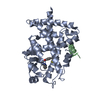
| ||||||||
|---|---|---|---|---|---|---|---|---|---|
| 1 |
| ||||||||
| Unit cell |
|
-Components
| #1: Protein | Mass: 31094.135 Da / Num. of mol.: 1 / Fragment: PPARgamma Ligand Binding Domain / Source method: isolated from a natural source / Source: (natural) Homo sapiens (human) / References: UniProt: P37231 |
|---|---|
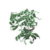
| #2: Protein/peptide | Mass: 1850.203 Da / Num. of mol.: 1 / Fragment: SHP 2nd LXXLL motif / Source method: isolated from a natural source / Source: (natural) Rattus norvegicus (Norway rat) / References: UniProt: P97947 |
| #3: Chemical | ChemComp-BRL / Rosiglitazone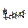  Mass: 357.427 Da / Num. of mol.: 1 / Source method: obtained synthetically / Formula: C18H19N3O3S Mass: 357.427 Da / Num. of mol.: 1 / Source method: obtained synthetically / Formula: C18H19N3O3S |
| #4: Water | ChemComp-HOH / Water Mass: 18.015 Da / Num. of mol.: 158 / Source method: isolated from a natural source / Formula: H2O Mass: 18.015 Da / Num. of mol.: 158 / Source method: isolated from a natural source / Formula: H2O |
-Experimental details
-Experiment
| Experiment | Method: X-RAY DIFFRACTION / Number of used crystals: 1 |
|---|
- Sample preparation
| Crystal | Density Matthews: 2.8 Å3/Da / Density % sol: 55.6 % |
|---|---|
| Crystal grow | Temperature: 293 K / Method: vapor diffusion, hanging drop / pH: 7 Details: PEG 3350, tri-sodium citrate, ethylene glycol, pH 7.0, VAPOR DIFFUSION, HANGING DROP, temperature 293K |
-Data collection
| Diffraction | Mean temperature: 100 K |
|---|---|
| Diffraction source | Source: SYNCHROTRON / Site: APS  / Beamline: 32-ID / Wavelength: 0.99998 Å / Beamline: 32-ID / Wavelength: 0.99998 Å |
| Detector | Type: MARRESEARCH / Detector: CCD / Date: Aug 5, 2004 |
| Radiation | Monochromator: Mirrors / Protocol: SINGLE WAVELENGTH / Monochromatic (M) / Laue (L): M / Scattering type: x-ray |
| Radiation wavelength | Wavelength: 0.99998 Å / Relative weight: 1 |
| Reflection | Resolution: 1.8→50 Å / Num. all: 33487 / Num. obs: 33433 / % possible obs: 100 % / Observed criterion σ(F): 32 / Observed criterion σ(I): 11165 / Biso Wilson estimate: 26.5 Å2 |
| Reflection shell | Resolution: 1.8→1.9 Å / % possible all: 99 |
- Processing
| Software |
| |||||||||||||||||||||||||
|---|---|---|---|---|---|---|---|---|---|---|---|---|---|---|---|---|---|---|---|---|---|---|---|---|---|---|
| Refinement | Method to determine structure: MOLECULAR REPLACEMENT Starting model: 1FM6 Resolution: 1.8→50 Å / Rfactor Rfree error: 0.005 / Data cutoff high absF: 1339168.9 / Data cutoff low absF: 0 / Isotropic thermal model: RESTRAINED / Cross valid method: THROUGHOUT / σ(F): 0 / Stereochemistry target values: Engh & Huber
| |||||||||||||||||||||||||
| Solvent computation | Solvent model: FLAT MODEL / Bsol: 61.248 Å2 / ksol: 0.370294 e/Å3 | |||||||||||||||||||||||||
| Displacement parameters | Biso mean: 46.7 Å2
| |||||||||||||||||||||||||
| Refine analyze |
| |||||||||||||||||||||||||
| Refinement step | Cycle: LAST / Resolution: 1.8→50 Å
| |||||||||||||||||||||||||
| Refine LS restraints |
| |||||||||||||||||||||||||
| LS refinement shell | Resolution: 1.8→1.91 Å / Rfactor Rfree error: 0.015 / Total num. of bins used: 6
| |||||||||||||||||||||||||
| Xplor file |
|
