 Movie
Movie Controller


+ Open data

- Basic information
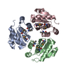
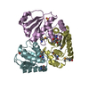
| Entry | Database: PDB / ID: 1zck | ||||||
|---|---|---|---|---|---|---|---|
| Title | native structure prl-1 (ptp4a1) | ||||||
 Components Components | protein tyrosine phosphatase 4a1 | ||||||
 Keywords Keywords |  HYDROLASE / prl-1 ptp4a1 HYDROLASE / prl-1 ptp4a1 | ||||||
| Function / homology |  Function and homology information Function and homology informationprotein tyrosine/serine/threonine phosphatase activity / dephosphorylation / protein-tyrosine-phosphatase / protein tyrosine phosphatase activity / cytoplasmic side of plasma membrane / spindle / early endosome / positive regulation of cell migration / cell cycle / endoplasmic reticulum ...protein tyrosine/serine/threonine phosphatase activity / dephosphorylation / protein-tyrosine-phosphatase / protein tyrosine phosphatase activity / cytoplasmic side of plasma membrane / spindle / early endosome / positive regulation of cell migration / cell cycle / endoplasmic reticulum / nucleus / cytoplasmSimilarity search - Function | ||||||
| Biological species |  Rattus norvegicus (Norway rat) Rattus norvegicus (Norway rat) | ||||||
| Method | X-RAY DIFFRACTION / SYNCHROTRON / MAD / Resolution: 1.9 Å | ||||||
 Authors Authors | Sun, J.P. / Wang, W.Q. / Yang, H. / Liu, S. / Liang, F. / Fedorov, A.A. / Almo, S.C. / Zhang, Z.Y. | ||||||
 Citation Citation | Journal: Biochemistry / Year: 2005 Title: Structure and Biochemical Properties of PRL-1, a Phosphatase Implicated in Cell Growth, Differentiation, and Tumor Invasion. Authors: Sun, J.P. / Wang, W.Q. / Yang, H. / Liu, S. / Liang, F. / Fedorov, A.A. / Almo, S.C. / Zhang, Z.Y. | ||||||
| History |
|
- Structure visualization
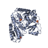
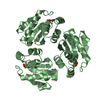
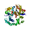
| Structure viewer | Molecule: MolmilJmol/JSmol |
|---|
- Downloads & links
-Download
| PDBx/mmCIF format | 1zck.cif.gz | 103.2 KB | Display | PDBx/mmCIF format |
|---|---|---|---|---|
| PDB format | pdb1zck.ent.gz | 85.4 KB | Display | PDB format |
| PDBx/mmJSON format | 1zck.json.gz | Tree view | PDBx/mmJSON format | |
| Others |  Other downloads Other downloads |
-Validation report
| Arichive directory | https://data.pdbj.org/pub/pdb/validation_reports/zc/1zckftp://data.pdbj.org/pub/pdb/validation_reports/zc/1zck | HTTPS FTP |
|---|
-Related structure data


-Links
 PDBj
PDBj


- Assembly
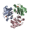
| Deposited unit | 
| ||||||||
|---|---|---|---|---|---|---|---|---|---|
| 1 |
| ||||||||
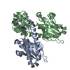
| Unit cell |
|
-Components
| #1: Protein | Mass: 17750.221 Da / Num. of mol.: 3 Source method: isolated from a genetically manipulated source Source: (gene. exp.) Rattus norvegicus (Norway rat) / Plasmid: pGEX / Production host:  Escherichia coli (E. coli) / Strain (production host): BL843 / References: UniProt: Q78EG7 Escherichia coli (E. coli) / Strain (production host): BL843 / References: UniProt: Q78EG7#2: Chemical | Acetic acid  Mass: 60.052 Da / Num. of mol.: 3 / Source method: obtained synthetically / Formula: C2H4O2 Mass: 60.052 Da / Num. of mol.: 3 / Source method: obtained synthetically / Formula: C2H4O2#3: Water | ChemComp-HOH / | Water Mass: 18.015 Da / Num. of mol.: 278 / Source method: isolated from a natural source / Formula: H2O Mass: 18.015 Da / Num. of mol.: 278 / Source method: isolated from a natural source / Formula: H2O |
|---|
-Experimental details
-Experiment
| Experiment | Method: X-RAY DIFFRACTION / Number of used crystals: 1 |
|---|
- Sample preparation
| Crystal | Density Matthews: 3.2 Å3/Da / Density % sol: 61.61 % |
|---|---|
| Crystal grow | Temperature: 277 K / Method: vapor diffusion, hanging drop / pH: 4.8 Details: Ammonium Sulfate, Sodium Acetate, pH 4.8, VAPOR DIFFUSION, HANGING DROP, temperature 277K |
-Data collection
| Diffraction | Mean temperature: 200 K | ||||||||||||
|---|---|---|---|---|---|---|---|---|---|---|---|---|---|
| Diffraction source | Source: SYNCHROTRON / Site: ALS  / Beamline: 5.0.1 / Wavelength: 0.9800, 0.97849, 0.96261 / Beamline: 5.0.1 / Wavelength: 0.9800, 0.97849, 0.96261 | ||||||||||||
| Detector | Type: RIGAKU RAXIS II / Detector: IMAGE PLATE / Date: Jun 30, 2003 | ||||||||||||
| Radiation | Monochromator: Si 111 CHANNEL / Protocol: MAD / Monochromatic (M) / Laue (L): M / Scattering type: x-ray | ||||||||||||
| Radiation wavelength |
| ||||||||||||
| Reflection | Resolution: 1.9→30 Å / Num. obs: 48966 / % possible obs: 96.9 % / Observed criterion σ(F): 0 / Observed criterion σ(I): 0 | ||||||||||||
| Reflection shell | Resolution: 1.9→1.97 Å / % possible all: 88.6 |
- Processing
| Software |
| ||||||||||||||||
|---|---|---|---|---|---|---|---|---|---|---|---|---|---|---|---|---|---|
| Refinement | Method to determine structure: MAD / Resolution: 1.9→30 Å / σ(F): 0 / Stereochemistry target values: Engh & Huber
| ||||||||||||||||
| Refinement step | Cycle: LAST / Resolution: 1.9→30 Å
|
