 Movie
Movie Controller


+ Open data

- Basic information
| Entry | Database: PDB / ID: 1xm2 | ||||||
|---|---|---|---|---|---|---|---|
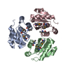
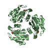
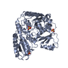
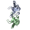
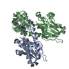
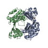
| Title | Crystal structure of Human PRL-1 | ||||||
 Components Components | Tyrosine Phosphatase Protein tyrosine phosphatase Protein tyrosine phosphatase | ||||||
 Keywords Keywords | HYDROLASE | ||||||
| Function / homology |  Function and homology information Function and homology informationprotein tyrosine/serine/threonine phosphatase activity / dephosphorylation / protein-tyrosine-phosphatase / protein tyrosine phosphatase activity / cytoplasmic side of plasma membrane / spindle / early endosome / positive regulation of cell migration / cell cycle / endoplasmic reticulum ...protein tyrosine/serine/threonine phosphatase activity / dephosphorylation / protein-tyrosine-phosphatase / protein tyrosine phosphatase activity / cytoplasmic side of plasma membrane / spindle / early endosome / positive regulation of cell migration / cell cycle / endoplasmic reticulum / nucleus / cytoplasmSimilarity search - Function | ||||||
| Biological species |  Homo sapiens (human) Homo sapiens (human) | ||||||
| Method | X-RAY DIFFRACTION / SYNCHROTRON / MAD / Resolution: 2.7 Å | ||||||
 Authors Authors | Jeong, D.G. / Kim, S.J. / Kim, J.H. / Son, J.H. / Ryu, S.E. | ||||||
 Citation Citation | Journal: J.Mol.Biol. / Year: 2005 Title: Trimeric structure of PRL-1 phosphatase reveals an active enzyme conformation and regulation mechanisms Authors: Jeong, D.G. / Kim, S.J. / Kim, J.H. / Son, J.H. / Park, M.R. / Lim, S.M. / Yoon, T.S. / Ryu, S.E. | ||||||
| History |
|
- Structure visualization
| Structure viewer | Molecule: MolmilJmol/JSmol |
|---|
- Downloads & links
-Download
| PDBx/mmCIF format | 1xm2.cif.gz | 181.2 KB | Display | PDBx/mmCIF format |
|---|---|---|---|---|
| PDB format | pdb1xm2.ent.gz | 152.8 KB | Display | PDB format |
| PDBx/mmJSON format | 1xm2.json.gz | Tree view | PDBx/mmJSON format | |
| Others |  Other downloads Other downloads |
-Validation report
| Arichive directory | https://data.pdbj.org/pub/pdb/validation_reports/xm/1xm2ftp://data.pdbj.org/pub/pdb/validation_reports/xm/1xm2 | HTTPS FTP |
|---|
-Related structure data
| Similar structure data |
|---|


-Links
 PDBj
PDBj


- Assembly
| Deposited unit | 
| ||||||||
|---|---|---|---|---|---|---|---|---|---|
| 1 | 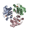
| ||||||||
| 2 | 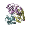
| ||||||||
| Unit cell |
| ||||||||
| Details | The biological assembly is a trimer. Two trimers exist in the asymmetric unit. |
-Components
| #1: Protein | Protein tyrosine phosphatase / PRL-1 Mass: 20063.527 Da / Num. of mol.: 6 / Mutation: C104S Source method: isolated from a genetically manipulated source Source: (gene. exp.) Homo sapiens (human) / Plasmid: pET28a / Production host:  Escherichia coli (E. coli) / References: UniProt: Q93096, protein-tyrosine-phosphatase Escherichia coli (E. coli) / References: UniProt: Q93096, protein-tyrosine-phosphatase#2: Chemical | ChemComp-SO4 / Sulfate  Mass: 96.063 Da / Num. of mol.: 6 / Source method: obtained synthetically / Formula: SO4 Mass: 96.063 Da / Num. of mol.: 6 / Source method: obtained synthetically / Formula: SO4#3: Water | ChemComp-HOH / | Water Mass: 18.015 Da / Num. of mol.: 92 / Source method: isolated from a natural source / Formula: H2O Mass: 18.015 Da / Num. of mol.: 92 / Source method: isolated from a natural source / Formula: H2O |
|---|
-Experimental details
-Experiment
| Experiment | Method: X-RAY DIFFRACTION / Number of used crystals: 1 |
|---|
- Sample preparation
| Crystal | Density Matthews: 2.51 Å3/Da / Density % sol: 51.06 % |
|---|---|
| Crystal grow | Temperature: 291 K / Method: vapor diffusion, hanging drop / pH: 7.5 Details: PEG 4K, Sodium Acetate, AMS, pH 7.5, VAPOR DIFFUSION, HANGING DROP, temperature 291K |
-Data collection
| Diffraction | Mean temperature: 100 K | |||||||||||||||
|---|---|---|---|---|---|---|---|---|---|---|---|---|---|---|---|---|
| Diffraction source | Source: SYNCHROTRON / Site: PAL/PLS  / Beamline: 6B / Wavelength: 0.979, 0.9792, 0.9794, 0.9716 / Beamline: 6B / Wavelength: 0.979, 0.9792, 0.9794, 0.9716 | |||||||||||||||
| Detector | Type: MACSCIENCE / Detector: IMAGE PLATE / Date: May 16, 2003 / Details: mirrors | |||||||||||||||
| Radiation | Monochromator: Mirrors / Protocol: MAD / Monochromatic (M) / Laue (L): M / Scattering type: x-ray | |||||||||||||||
| Radiation wavelength |
| |||||||||||||||
| Reflection | Resolution: 2.7→40 Å / Num. all: 32950 / Num. obs: 31124 / % possible obs: 94.5 % / Observed criterion σ(F): 0 / Observed criterion σ(I): 2 / Redundancy: 4.3 % / Rmerge(I) obs: 0.079 / Rsym value: 0.079 / Net I/σ(I): 7.2 | |||||||||||||||
| Reflection shell | Resolution: 2.7→2.85 Å / Redundancy: 4.3 % / Rmerge(I) obs: 0.26 / Mean I/σ(I) obs: 2.9 / Num. unique all: 4531 / Rsym value: 0.26 / % possible all: 95.4 |
- Processing
| Software |
| |||||||||||||||||||||||||
|---|---|---|---|---|---|---|---|---|---|---|---|---|---|---|---|---|---|---|---|---|---|---|---|---|---|---|
| Refinement | Method to determine structure: MAD / Resolution: 2.7→40 Å / Isotropic thermal model: isotropic / Cross valid method: THROUGHOUT / σ(F): 0 / σ(I): 0 / Stereochemistry target values: Engh & Huber
| |||||||||||||||||||||||||
| Refinement step | Cycle: LAST / Resolution: 2.7→40 Å
| |||||||||||||||||||||||||
| Refine LS restraints |
|
