 Movie
Movie Controller

[English] 日本語
 Yorodumi
- PDB-1z9o: 1.9 Angstrom Crystal Structure of the Rat VAP-A MSP Homology Doma... -
+ Open data

- Basic information
| Entry | Database: PDB / ID: 1z9o | ||||||
|---|---|---|---|---|---|---|---|
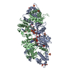
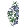
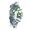
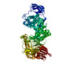
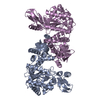
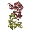
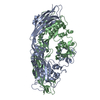
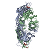
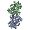
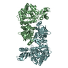
| Title | 1.9 Angstrom Crystal Structure of the Rat VAP-A MSP Homology Domain in Complex with the Rat ORP1 FFAT Motif | ||||||
 Components Components |
| ||||||
 Keywords Keywords | PROTEIN BINDING/LIPID BINDING PROTEIN / VAP-33 / VAP-A / VAP-B / VAP-C / VAPA /  VAPB / VAPC / VAP-33a / Scs2 / ERG30 / FFAT motif / ER / endoplasmic reticulum / targeting / immunoglobulin-like beta sheet / MSP homology domain / OSBP / ORP / oxysterol binding protein / PROTEIN BINDING-LIPID BINDING PROTEIN COMPLEX VAPB / VAPC / VAP-33a / Scs2 / ERG30 / FFAT motif / ER / endoplasmic reticulum / targeting / immunoglobulin-like beta sheet / MSP homology domain / OSBP / ORP / oxysterol binding protein / PROTEIN BINDING-LIPID BINDING PROTEIN COMPLEX | ||||||
| Function / homology |  Function and homology information Function and homology informationSynthesis of bile acids and bile salts / FFAT motif binding / endoplasmic reticulum-plasma membrane tethering / organelle membrane contact site / ceramide transport / protein localization to endoplasmic reticulum / sphingomyelin biosynthetic process / : / sterol transport / sterol binding ...Synthesis of bile acids and bile salts / FFAT motif binding / endoplasmic reticulum-plasma membrane tethering / organelle membrane contact site / ceramide transport / protein localization to endoplasmic reticulum / sphingomyelin biosynthetic process / : / sterol transport / sterol binding / positive regulation by host of viral genome replication / COPII-coated vesicle budding / MHC class II antigen presentation / negative regulation by host of viral genome replication / Insertion of tail-anchored proteins into the endoplasmic reticulum membrane / phospholipid transport / cholesterol transport / Neutrophil degranulation / cholesterol binding / viral release from host cell / bicellular tight junction / endoplasmic reticulum to Golgi vesicle-mediated transport / neuron projection development / microtubule cytoskeleton / late endosome / microtubule binding / nuclear membrane / vesicle / endosome / protein heterodimerization activity / protein domain specific binding / Golgi membrane / intracellular membrane-bounded organelle / protein-containing complex binding / endoplasmic reticulum membrane / endoplasmic reticulum / protein homodimerization activity / membrane / identical protein binding / plasma membrane / cytosolSimilarity search - Function | ||||||
| Biological species |  Rattus norvegicus (Norway rat) Rattus norvegicus (Norway rat) | ||||||
| Method | X-RAY DIFFRACTION / SYNCHROTRON / SAD / Resolution: 1.9 Å | ||||||
 Authors Authors | Kaiser, S.E. / Brickner, J.H. / Reilein, A.R. / Fenn, T.D. / Walter, P. / Brunger, A.T. | ||||||
 Citation Citation | Journal: Structure / Year: 2005 Title: Structural basis of FFAT motif-mediated ER targeting Authors: Kaiser, S.E. / Brickner, J.H. / Reilein, A.R. / Fenn, T.D. / Walter, P. / Brunger, A.T. | ||||||
| History |
|
- Structure visualization
| Structure viewer | Molecule: MolmilJmol/JSmol |
|---|
- Downloads & links
-Download
| PDBx/mmCIF format | 1z9o.cif.gz | 166 KB | Display | PDBx/mmCIF format |
|---|---|---|---|---|
| PDB format | pdb1z9o.ent.gz | 132.8 KB | Display | PDB format |
| PDBx/mmJSON format | 1z9o.json.gz | Tree view | PDBx/mmJSON format | |
| Others |  Other downloads Other downloads |
-Validation report
| Arichive directory | https://data.pdbj.org/pub/pdb/validation_reports/z9/1z9oftp://data.pdbj.org/pub/pdb/validation_reports/z9/1z9o | HTTPS FTP |
|---|
-Related structure data
| Related structure data | 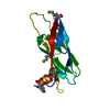 1z9lSC S: Starting model for refinement C: citing same article ( |
|---|---|
| Similar structure data |
-Links
 PDBj
PDBj


- Assembly
| Deposited unit | 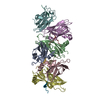
| ||||||||||||||||||||||||||||||||||||||||||||||||||||||||||
|---|---|---|---|---|---|---|---|---|---|---|---|---|---|---|---|---|---|---|---|---|---|---|---|---|---|---|---|---|---|---|---|---|---|---|---|---|---|---|---|---|---|---|---|---|---|---|---|---|---|---|---|---|---|---|---|---|---|---|---|
| 1 |
| ||||||||||||||||||||||||||||||||||||||||||||||||||||||||||
| Unit cell |
| ||||||||||||||||||||||||||||||||||||||||||||||||||||||||||
| Noncrystallographic symmetry (NCS) | NCS domain:
NCS domain segments: Component-ID: 1 / Beg auth comp-ID: GLU / Beg label comp-ID: GLU / Refine code: 1
NCS ensembles :
|
-Components
| #1: Protein | Mass: 14545.880 Da / Num. of mol.: 6 Source method: isolated from a genetically manipulated source Source: (gene. exp.) Rattus norvegicus (Norway rat) / Gene: Vapa, Vap33 / Plasmid: pGEX / Species (production host): Escherichia coli / Production host:  Escherichia coli BL21 (bacteria) / Strain (production host): BL21 / References: GenBank: 61889097, UniProt: Q9Z270*PLUS Escherichia coli BL21 (bacteria) / Strain (production host): BL21 / References: GenBank: 61889097, UniProt: Q9Z270*PLUS#2: Protein/peptide | Oxysterol-binding protein / ORP-1Mass: 1175.157 Da / Num. of mol.: 6 / Fragment: Residues 472-481 / Source method: obtained synthetically Details: peptide NH2-SEDEFYDALS-COO corresponding to rat ORP1 472-481 synthesized by Biopeptide LLC References: UniProt: Q8K4M9 #3: Water | ChemComp-HOH / | Water Mass: 18.015 Da / Num. of mol.: 185 / Source method: isolated from a natural source / Formula: H2O Mass: 18.015 Da / Num. of mol.: 185 / Source method: isolated from a natural source / Formula: H2O |
|---|
-Experimental details
-Experiment
| Experiment | Method: X-RAY DIFFRACTION / Number of used crystals: 2 |
|---|
- Sample preparation
| Crystal | Density Matthews: 2.12 Å3/Da / Density % sol: 41 % |
|---|---|
| Crystal grow | Temperature: 283 K / Method: vapor diffusion, sitting drop / pH: 7.4 Details: PEG 2000 MME, sodium thiocyanate, tris buffer, pH 7.4, VAPOR DIFFUSION, SITTING DROP, temperature 283K |
-Data collection
| Diffraction |
| ||||||||||||||||||
|---|---|---|---|---|---|---|---|---|---|---|---|---|---|---|---|---|---|---|---|
| Diffraction source |
| ||||||||||||||||||
| Detector |
| ||||||||||||||||||
| Radiation |
| ||||||||||||||||||
| Radiation wavelength |
| ||||||||||||||||||
| Reflection | Resolution: 1.9→50 Å / Num. all: 59859 / Num. obs: 59506 / Observed criterion σ(F): 1 / Observed criterion σ(I): 1 / Redundancy: 4 % | ||||||||||||||||||
| Reflection shell | Resolution: 1.9→1.949 Å / % possible all: 100 |

- Processing
| Software |
| |||||||||||||||||||||||||||||||||||||||||||||||||||||||||||||||||||||||||||||||||||||||||||||||||||||||||||||||||||||||||||||||||||||||||||||||||||||||||||||||||||||||||||||||
|---|---|---|---|---|---|---|---|---|---|---|---|---|---|---|---|---|---|---|---|---|---|---|---|---|---|---|---|---|---|---|---|---|---|---|---|---|---|---|---|---|---|---|---|---|---|---|---|---|---|---|---|---|---|---|---|---|---|---|---|---|---|---|---|---|---|---|---|---|---|---|---|---|---|---|---|---|---|---|---|---|---|---|---|---|---|---|---|---|---|---|---|---|---|---|---|---|---|---|---|---|---|---|---|---|---|---|---|---|---|---|---|---|---|---|---|---|---|---|---|---|---|---|---|---|---|---|---|---|---|---|---|---|---|---|---|---|---|---|---|---|---|---|---|---|---|---|---|---|---|---|---|---|---|---|---|---|---|---|---|---|---|---|---|---|---|---|---|---|---|---|---|---|---|---|---|---|
| Refinement | Method to determine structure: SAD Starting model: Found Se by molecular replacement w/ 1Z9L Resolution: 1.9→14.65 Å / Cor.coef. Fo:Fc: 0.95 / Cor.coef. Fo:Fc free: 0.928 / SU B: 10.431 / SU ML: 0.149 / TLS residual ADP flag: LIKELY RESIDUAL / Cross valid method: THROUGHOUT / ESU R: 0.199 / ESU R Free: 0.176 / Stereochemistry target values: MAXIMUM LIKELIHOOD / Details: HYDROGENS HAVE BEEN ADDED IN THE RIDING POSITIONS
| |||||||||||||||||||||||||||||||||||||||||||||||||||||||||||||||||||||||||||||||||||||||||||||||||||||||||||||||||||||||||||||||||||||||||||||||||||||||||||||||||||||||||||||||
| Solvent computation | Ion probe radii: 0.8 Å / Shrinkage radii: 0.8 Å / VDW probe radii: 1.2 Å / Solvent model: MASK | |||||||||||||||||||||||||||||||||||||||||||||||||||||||||||||||||||||||||||||||||||||||||||||||||||||||||||||||||||||||||||||||||||||||||||||||||||||||||||||||||||||||||||||||
| Displacement parameters | Biso mean: 26.342 Å2
| |||||||||||||||||||||||||||||||||||||||||||||||||||||||||||||||||||||||||||||||||||||||||||||||||||||||||||||||||||||||||||||||||||||||||||||||||||||||||||||||||||||||||||||||
| Refinement step | Cycle: LAST / Resolution: 1.9→14.65 Å
| |||||||||||||||||||||||||||||||||||||||||||||||||||||||||||||||||||||||||||||||||||||||||||||||||||||||||||||||||||||||||||||||||||||||||||||||||||||||||||||||||||||||||||||||
| Refine LS restraints |
| |||||||||||||||||||||||||||||||||||||||||||||||||||||||||||||||||||||||||||||||||||||||||||||||||||||||||||||||||||||||||||||||||||||||||||||||||||||||||||||||||||||||||||||||
| Refine LS restraints NCS | Dom-ID: 1 / Refine-ID: X-RAY DIFFRACTION
| |||||||||||||||||||||||||||||||||||||||||||||||||||||||||||||||||||||||||||||||||||||||||||||||||||||||||||||||||||||||||||||||||||||||||||||||||||||||||||||||||||||||||||||||
| LS refinement shell | Resolution: 1.9→1.949 Å / Total num. of bins used: 20
| |||||||||||||||||||||||||||||||||||||||||||||||||||||||||||||||||||||||||||||||||||||||||||||||||||||||||||||||||||||||||||||||||||||||||||||||||||||||||||||||||||||||||||||||
| Refinement TLS params. | Method: refined / Refine-ID: X-RAY DIFFRACTION
| |||||||||||||||||||||||||||||||||||||||||||||||||||||||||||||||||||||||||||||||||||||||||||||||||||||||||||||||||||||||||||||||||||||||||||||||||||||||||||||||||||||||||||||||
| Refinement TLS group |
|
