- PDB-1z9k: Photosynthetic Reaction Center from Rhodobacter sphaeroides -
+
Open data
ID or keywords:
Loading...
-
Basic information
Entry
Database: PDB / ID: 1z9k
Title
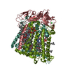
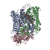
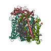
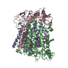
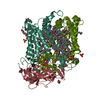
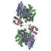
Photosynthetic Reaction Center from Rhodobacter sphaeroides
Components
(Reaction center protein ...Photosynthetic reaction centre) x 3
Keywords
PHOTOSYNTHESIS / ALPHA HELIX / MEMBRANE PROTEIN
Function / homology
Function and homology information
: / plasma membrane-derived chromatophore membrane / plasma membrane light-harvesting complex / bacteriochlorophyll binding / photosynthesis, light reaction / electron transporter, transferring electrons within the cyclic electron transport pathway of photosynthesis activity / photosynthetic electron transport in photosystem II / membrane => GO:0016020 / metal ion binding Similarity search - Function
Photosynthetic reaction centre, H subunit / Bacterial photosynthetic reaction centre, H-chain, C-terminal / Photosynthetic reaction centre, M subunit / Photosynthetic reaction centre, H subunit, N-terminal / Photosynthetic reaction centre, H subunit, N-terminal domain superfamily / Photosynthetic reaction centre, H-chain N-terminal region / PRC-barrel domain / PRC-barrel domain / Photosynthetic reaction centre, L subunit / PRC-barrel-like superfamily ...Photosynthetic reaction centre, H subunit / Bacterial photosynthetic reaction centre, H-chain, C-terminal / Photosynthetic reaction centre, M subunit / Photosynthetic reaction centre, H subunit, N-terminal / Photosynthetic reaction centre, H subunit, N-terminal domain superfamily / Photosynthetic reaction centre, H-chain N-terminal region / PRC-barrel domain / PRC-barrel domain / Photosynthetic reaction centre, L subunit / PRC-barrel-like superfamily / Photosynthetic reaction centre, L/M / Photosystem II protein D1/D2 superfamily / Photosynthetic reaction centre protein / Photosynthetic reaction center proteins signature. Similarity search - Domain/homology
BACTERIOCHLOROPHYLL A / BACTERIOPHEOPHYTIN A / : / : / UBIQUINONE-10 / Reaction center protein M chain / Reaction center protein L chain / Reaction center protein H chain / Reaction center protein L chain / Reaction center protein M chain / Reaction center protein H chain Similarity search - Component
Biological species
Rhodobacter sphaeroides (bacteria)
Method
X-RAY DIFFRACTION / MOLECULAR REPLACEMENT / Resolution: 4.6 Å
In the structure databanks used in Yorodumi, some data are registered as the other names, "COVID-19 virus" and "2019-nCoV". Here are the details of the virus and the list of structure data.
Jan 31, 2019. EMDB accession codes are about to change! (news from PDBe EMDB page)
EMDB accession codes are about to change! (news from PDBe EMDB page)
The allocation of 4 digits for EMDB accession codes will soon come to an end. Whilst these codes will remain in use, new EMDB accession codes will include an additional digit and will expand incrementally as the available range of codes is exhausted. The current 4-digit format prefixed with “EMD-” (i.e. EMD-XXXX) will advance to a 5-digit format (i.e. EMD-XXXXX), and so on. It is currently estimated that the 4-digit codes will be depleted around Spring 2019, at which point the 5-digit format will come into force.
The EM Navigator/Yorodumi systems omit the EMD- prefix.
Related info.:Q: What is EMD? / ID/Accession-code notation in Yorodumi/EM Navigator
Yorodumi is a browser for structure data from EMDB, PDB, SASBDB, etc.
This page is also the successor to EM Navigator detail page, and also detail information page/front-end page for Omokage search.
The word "yorodu" (or yorozu) is an old Japanese word meaning "ten thousand". "mi" (miru) is to see.
Related info.:EMDB / PDB / SASBDB / Comparison of 3 databanks / Yorodumi Search / Aug 31, 2016. New EM Navigator & Yorodumi / Yorodumi Papers / Jmol/JSmol / Function and homology information / Changes in new EM Navigator and Yorodumi
 Movie
Movie Controller


 Open data
 Basic information
 Components
 Photosynthetic reaction centre) x 3
  Keywords
 Function and homology information
 Authors
 Citation
 Structure visualization
 Downloads & links
 Other downloads


 PDBj
PDBj


 Assembly
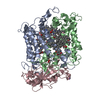


 Mass: 911.504 Da / Num. of mol.: 4 / Source method: obtained synthetically / Formula: C55H74MgN4O6
 Mass: 889.215 Da / Num. of mol.: 2 / Source method: obtained synthetically / Formula: C55H76N4O6
 Mass: 863.343 Da / Num. of mol.: 2 / Source method: obtained synthetically / Formula: C59H90O4
 Mass: 55.845 Da / Num. of mol.: 1 / Source method: obtained synthetically / Formula: Fe
 Mass: 54.938 Da / Num. of mol.: 1 / Source method: obtained synthetically / Formula: Mn
 Sample preparation
 Processing