 Movie
Movie Controller


+ Open data

- Basic information
| Entry | Database: PDB / ID: 1z7g | ||||||
|---|---|---|---|---|---|---|---|
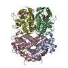
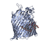
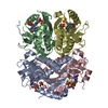
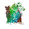
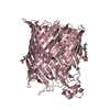
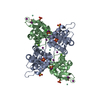
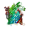
| Title | Free human HGPRT | ||||||
 Components Components | Hypoxanthine-guanine phosphoribosyltransferase | ||||||
 Keywords Keywords | TRANSFERASE / flexibility / trans cis peptide bond isomerization / nucleotide binding | ||||||
| Function / homology |  Function and homology information Function and homology informationadenine metabolic process / Defective HPRT1 disrupts guanine and hypoxanthine salvage / GMP catabolic process / guanine salvage / hypoxanthine metabolic process / hypoxanthine salvage / cerebral cortex neuron differentiation / positive regulation of dopamine metabolic process / lymphocyte proliferation / hypoxanthine phosphoribosyltransferase ...adenine metabolic process / Defective HPRT1 disrupts guanine and hypoxanthine salvage / GMP catabolic process / guanine salvage / hypoxanthine metabolic process / hypoxanthine salvage / cerebral cortex neuron differentiation / positive regulation of dopamine metabolic process / lymphocyte proliferation / hypoxanthine phosphoribosyltransferase / IMP metabolic process / GMP salvage / guanine phosphoribosyltransferase activity / hypoxanthine phosphoribosyltransferase activity / grooming behavior / IMP salvage / Purine salvage / striatum development / AMP salvage / dopaminergic neuron differentiation / purine nucleotide biosynthetic process / Azathioprine ADME / purine ribonucleoside salvage / dendrite morphogenesis / dopamine metabolic process / central nervous system neuron development / response to amphetamine / locomotory behavior / T cell mediated cytotoxicity / protein homotetramerization / nucleotide binding / magnesium ion binding / extracellular exosome / identical protein binding / cytosol / cytoplasmSimilarity search - Function | ||||||
| Biological species |  Homo sapiens (human) Homo sapiens (human) | ||||||
| Method | X-RAY DIFFRACTION / MOLECULAR REPLACEMENT / Resolution: 1.9 Å | ||||||
 Authors Authors | Keough, D.T. / Brereton, I.M. / de Jersey, J. / Guddat, L.W. | ||||||
 Citation Citation | Journal: J.Mol.Biol. / Year: 2005 Title: The Crystal Structure of Free Human Hypoxanthine-guanine Phosphoribosyltransferase Reveals Extensive Conformational Plasticity Throughout the Catalytic Cycle Authors: Keough, D.T. / Brereton, I.M. / de Jersey, J. / Guddat, L.W. | ||||||
| History |
|
- Structure visualization
| Structure viewer | Molecule: MolmilJmol/JSmol |
|---|
- Downloads & links
-Download
| PDBx/mmCIF format | 1z7g.cif.gz | 176.1 KB | Display | PDBx/mmCIF format |
|---|---|---|---|---|
| PDB format | pdb1z7g.ent.gz | 139.7 KB | Display | PDB format |
| PDBx/mmJSON format | 1z7g.json.gz | Tree view | PDBx/mmJSON format | |
| Others |  Other downloads Other downloads |
-Validation report
| Arichive directory | https://data.pdbj.org/pub/pdb/validation_reports/z7/1z7gftp://data.pdbj.org/pub/pdb/validation_reports/z7/1z7g | HTTPS FTP |
|---|
-Related structure data
| Related structure data | 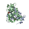 1hmpS S: Starting model for refinement |
|---|---|
| Similar structure data |


-Links
 PDBj
PDBj
- Assembly
| Deposited unit | 
| ||||||||
|---|---|---|---|---|---|---|---|---|---|
| 1 | 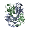
| ||||||||
| 2 | 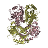
| ||||||||
| Unit cell |
| ||||||||
| Details | The biological unit is a tetramer. The asymmetric unit consists of two seperate dimers. Tetramers are generated by crystallographic two fold axes of symmetry. |
-Components
| #1: Protein | / HGPRT / HGPRTase Mass: 24481.217 Da / Num. of mol.: 4 Source method: isolated from a genetically manipulated source Source: (gene. exp.) Homo sapiens (human) / Gene: HPRT1, HPRTPlasmid details: recombinant plasmid pHTM was obtained by site directed mutagenesis of pRG1 which in turn is derived from pT7-7 Plasmid: pHTM / Production host:  Escherichia coli (E. coli) / Strain (production host): S 606 Escherichia coli (E. coli) / Strain (production host): S 606References: UniProt: P00492, hypoxanthine phosphoribosyltransferase#2: Water | ChemComp-HOH / | Water Mass: 18.015 Da / Num. of mol.: 504 / Source method: isolated from a natural source / Formula: H2O Mass: 18.015 Da / Num. of mol.: 504 / Source method: isolated from a natural source / Formula: H2O |
|---|
-Experimental details
-Experiment
| Experiment | Method: X-RAY DIFFRACTION / Number of used crystals: 1 |
|---|
- Sample preparation
| Crystal | Density Matthews: 2.1 Å3/Da / Density % sol: 39.1 % |
|---|---|
| Crystal grow | Temperature: 291 K / Method: vapor diffusion, hanging drop / pH: 4.6 Details: 0.2 M ammonium acetate, 0.1 M sodium acetate, 30% PEG4000, pH 4.6, VAPOR DIFFUSION, HANGING DROP, temperature 291K |
-Data collection
| Diffraction | Mean temperature: 100 K |
|---|---|
| Diffraction source | Source: ROTATING ANODE / Type: RIGAKU FR-E / Wavelength: 1.5418 Å |
| Detector | Type: RIGAKU RAXIS IV / Detector: IMAGE PLATE / Date: Jun 3, 2003 / Details: Osmic |
| Radiation | Monochromator: Single wavelength / Protocol: SINGLE WAVELENGTH / Monochromatic (M) / Laue (L): M / Scattering type: x-ray |
| Radiation wavelength | Wavelength: 1.5418 Å / Relative weight: 1 |
| Reflection | Resolution: 1.9→46.3 Å / Num. all: 66957 / Num. obs: 66957 / % possible obs: 95.6 % / Observed criterion σ(F): 0 / Observed criterion σ(I): 0 / Redundancy: 2.5 % / Rmerge(I) obs: 0.062 / Rsym value: 0.062 / Net I/σ(I): 12.3 |
| Reflection shell | Resolution: 1.9→1.97 Å / Redundancy: 1.7 % / Rmerge(I) obs: 0.288 / Mean I/σ(I) obs: 2.2 / Num. unique all: 5419 / Rsym value: 0.288 / % possible all: 79.3 |
- Processing
| Software |
| |||||||||||||||||||||||||
|---|---|---|---|---|---|---|---|---|---|---|---|---|---|---|---|---|---|---|---|---|---|---|---|---|---|---|
| Refinement | Method to determine structure: MOLECULAR REPLACEMENT Starting model: PDB ENTRY 1HMP Resolution: 1.9→46.3 Å / Isotropic thermal model: Isotropic / Cross valid method: THROUGHOUT / σ(F): 1 / Stereochemistry target values: Engh & Huber
| |||||||||||||||||||||||||
| Displacement parameters | Biso mean: 59.11 Å2 | |||||||||||||||||||||||||
| Refinement step | Cycle: LAST / Resolution: 1.9→46.3 Å
| |||||||||||||||||||||||||
| Refine LS restraints |
| |||||||||||||||||||||||||
| LS refinement shell | Resolution: 1.9→1.97 Å / Rfactor Rfree error: 0.034
| |||||||||||||||||||||||||
| Xplor file |
|
