 Movie
Movie Controller


+ Open data

- Basic information
| Entry | Database: PDB / ID: 1z0a | ||||||
|---|---|---|---|---|---|---|---|
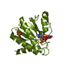
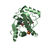
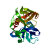
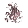
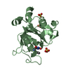
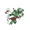
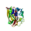
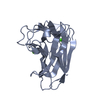
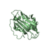
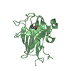
| Title | GDP-Bound Rab2A GTPase | ||||||
 Components Components | Ras-related protein Rab-2A | ||||||
 Keywords Keywords |  PROTEIN TRANSPORT / Rab GTPase / Rab2 / Vesicular trafficking PROTEIN TRANSPORT / Rab GTPase / Rab2 / Vesicular trafficking | ||||||
| Function / homology |  Function and homology information Function and homology informationRAB geranylgeranylation / Golgi Cisternae Pericentriolar Stack Reorganization / Golgi organization / endoplasmic reticulum to Golgi vesicle-mediated transport / endoplasmic reticulum-Golgi intermediate compartment membrane / post-translational protein modification / GDP binding / melanosome / protein transport / lysosomal membrane ...RAB geranylgeranylation / Golgi Cisternae Pericentriolar Stack Reorganization / Golgi organization / endoplasmic reticulum to Golgi vesicle-mediated transport / endoplasmic reticulum-Golgi intermediate compartment membrane / post-translational protein modification / GDP binding / melanosome / protein transport / lysosomal membrane / Golgi membrane / GTPase activity / endoplasmic reticulum membrane / GTP binding / Golgi apparatus / extracellular exosome / nucleus / cytosolSimilarity search - Function | ||||||
| Biological species |  Homo sapiens (human) Homo sapiens (human) | ||||||
| Method | X-RAY DIFFRACTION / SYNCHROTRON / MOLECULAR REPLACEMENT / Resolution: 2.12 Å | ||||||
 Authors Authors | Eathiraj, S. / Pan, X. / Ritacco, C. / Lambright, D.G. | ||||||
 Citation Citation | Journal: Nature / Year: 2005 Title: Structural basis of family-wide Rab GTPase recognition by rabenosyn-5. Authors: Eathiraj, S. / Pan, X. / Ritacco, C. / Lambright, D.G. | ||||||
| History |
|
- Structure visualization
| Structure viewer | Molecule: MolmilJmol/JSmol |
|---|
- Downloads & links
-Download
| PDBx/mmCIF format | 1z0a.cif.gz | 155 KB | Display | PDBx/mmCIF format |
|---|---|---|---|---|
| PDB format | pdb1z0a.ent.gz | 119.5 KB | Display | PDB format |
| PDBx/mmJSON format | 1z0a.json.gz | Tree view | PDBx/mmJSON format | |
| Others |  Other downloads Other downloads |
-Validation report
| Arichive directory | https://data.pdbj.org/pub/pdb/validation_reports/z0/1z0aftp://data.pdbj.org/pub/pdb/validation_reports/z0/1z0a | HTTPS FTP |
|---|
-Related structure data
| Related structure data | 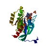 1yu9C 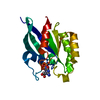 1yvdC 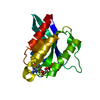 1yzkC 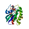 1yzlC 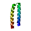 1yzmC 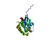 1yznC 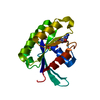 1yzqC 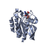 1yztC 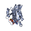 1yzuC 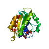 1z06C 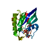 1z07C 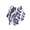 1z08C 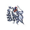 1z0dC 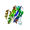 1z0fC 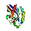 1z0iC 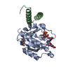 1z0jC  1z0kC 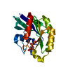 1z22C 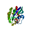 1z2aC C: citing same article ( |
|---|---|
| Similar structure data |
-Links
 PDBj
PDBj- Assembly
| Deposited unit | 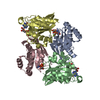
| ||||||||
|---|---|---|---|---|---|---|---|---|---|
| 1 | 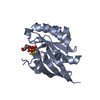
| ||||||||
| 2 | 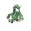
| ||||||||
| 3 | 
| ||||||||
| 4 | 
| ||||||||
| 5 |
| ||||||||
| 6 | 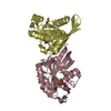
| ||||||||
| 7 | 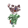
| ||||||||
| Unit cell |
|
-Components
| #1: Protein | Mass: 19720.361 Da / Num. of mol.: 4 Source method: isolated from a genetically manipulated source Source: (gene. exp.) Homo sapiens (human) / Gene: RAB2, RAB2A / Plasmid: pGEX6P1 / Production host:  Escherichia coli (E. coli) / Strain (production host): BL21 (DE3) Codon Plus-RIL Cells / References: UniProt: P61019 Escherichia coli (E. coli) / Strain (production host): BL21 (DE3) Codon Plus-RIL Cells / References: UniProt: P61019#2: Chemical | ChemComp-MG /   Mass: 24.305 Da / Num. of mol.: 4 / Source method: obtained synthetically / Formula: Mg Mass: 24.305 Da / Num. of mol.: 4 / Source method: obtained synthetically / Formula: Mg#3: Chemical | ChemComp-GDP / Guanosine diphosphate  Type: RNA linking / Mass: 443.201 Da / Num. of mol.: 4 / Source method: obtained synthetically / Formula: C10H15N5O11P2 / Comment: GDP, energy-carrying molecule*YM Type: RNA linking / Mass: 443.201 Da / Num. of mol.: 4 / Source method: obtained synthetically / Formula: C10H15N5O11P2 / Comment: GDP, energy-carrying molecule*YM#4: Water | ChemComp-HOH / | Water Mass: 18.015 Da / Num. of mol.: 533 / Source method: isolated from a natural source / Formula: H2O Mass: 18.015 Da / Num. of mol.: 533 / Source method: isolated from a natural source / Formula: H2O |
|---|
-Experimental details
-Experiment
| Experiment | Method: X-RAY DIFFRACTION / Number of used crystals: 1 |
|---|
- Sample preparation
| Crystal | Density Matthews: 3 Å3/Da / Density % sol: 58.71 % |
|---|---|
| Crystal grow | Temperature: 291 K / Method: vapor diffusion, hanging drop / pH: 5.5 Details: 15% PEG 4000, 400mM Lithium sulfate, 100mM Citrate, pH 5.5, VAPOR DIFFUSION, HANGING DROP, temperature 291K |
-Data collection
| Diffraction | Mean temperature: 100 K |
|---|---|
| Diffraction source | Source: SYNCHROTRON / Site: NSLS  / Beamline: X25 / Wavelength: 1.1 Å / Beamline: X25 / Wavelength: 1.1 Å |
| Detector | Type: ADSC QUANTUM 315 / Detector: CCD / Date: Sep 26, 2004 |
| Radiation | Monochromator: Si(111) / Protocol: SINGLE WAVELENGTH / Monochromatic (M) / Laue (L): M / Scattering type: x-ray |
| Radiation wavelength | Wavelength: 1.1 Å / Relative weight: 1 |
| Reflection | Resolution: 2.12→50 Å / Num. obs: 49343 / % possible obs: 68.5 % / Observed criterion σ(F): 0 / Observed criterion σ(I): 0 / Redundancy: 4 % / Biso Wilson estimate: 45.2 Å2 / Rsym value: 0.069 / Net I/σ(I): 32.9 |
| Reflection shell | Resolution: 2.12→2.18 Å / Redundancy: 5 % / Mean I/σ(I) obs: 3.7 / Num. unique all: 1880 / Rsym value: 0.377 / % possible all: 45.8 |
- Processing
| Software |
| ||||||||||||||||||||||||||||||||||||||||||||||||||||||||||||||||||||||||||||||||||||||||||||||||||||||||||||||||||||||||||||||||||||||||||||||||||||||||||||||||||||||||||
|---|---|---|---|---|---|---|---|---|---|---|---|---|---|---|---|---|---|---|---|---|---|---|---|---|---|---|---|---|---|---|---|---|---|---|---|---|---|---|---|---|---|---|---|---|---|---|---|---|---|---|---|---|---|---|---|---|---|---|---|---|---|---|---|---|---|---|---|---|---|---|---|---|---|---|---|---|---|---|---|---|---|---|---|---|---|---|---|---|---|---|---|---|---|---|---|---|---|---|---|---|---|---|---|---|---|---|---|---|---|---|---|---|---|---|---|---|---|---|---|---|---|---|---|---|---|---|---|---|---|---|---|---|---|---|---|---|---|---|---|---|---|---|---|---|---|---|---|---|---|---|---|---|---|---|---|---|---|---|---|---|---|---|---|---|---|---|---|---|---|---|---|
| Refinement | Method to determine structure: MOLECULAR REPLACEMENT Starting model: Polyalanine Rab4 GTPase Resolution: 2.12→10 Å / Cor.coef. Fo:Fc: 0.948 / Cor.coef. Fo:Fc free: 0.913 / SU B: 5.888 / SU ML: 0.156 / Cross valid method: THROUGHOUT / σ(F): 0 / σ(I): 0 / ESU R: 0.208 / ESU R Free: 0.211 / Stereochemistry target values: MAXIMUM LIKELIHOOD / Details: HYDROGENS HAVE BEEN ADDED IN THE RIDING POSITIONS
| ||||||||||||||||||||||||||||||||||||||||||||||||||||||||||||||||||||||||||||||||||||||||||||||||||||||||||||||||||||||||||||||||||||||||||||||||||||||||||||||||||||||||||
| Solvent computation | Ion probe radii: 0.8 Å / Shrinkage radii: 0.8 Å / VDW probe radii: 1.2 Å / Solvent model: MASK | ||||||||||||||||||||||||||||||||||||||||||||||||||||||||||||||||||||||||||||||||||||||||||||||||||||||||||||||||||||||||||||||||||||||||||||||||||||||||||||||||||||||||||
| Displacement parameters | Biso mean: 44.912 Å2
| ||||||||||||||||||||||||||||||||||||||||||||||||||||||||||||||||||||||||||||||||||||||||||||||||||||||||||||||||||||||||||||||||||||||||||||||||||||||||||||||||||||||||||
| Refinement step | Cycle: LAST / Resolution: 2.12→10 Å
| ||||||||||||||||||||||||||||||||||||||||||||||||||||||||||||||||||||||||||||||||||||||||||||||||||||||||||||||||||||||||||||||||||||||||||||||||||||||||||||||||||||||||||
| Refine LS restraints |
| ||||||||||||||||||||||||||||||||||||||||||||||||||||||||||||||||||||||||||||||||||||||||||||||||||||||||||||||||||||||||||||||||||||||||||||||||||||||||||||||||||||||||||
| LS refinement shell | Resolution: 2.12→2.173 Å / Total num. of bins used: 20
|
