 Movie
Movie Controller

[English] 日本語
 Yorodumi
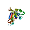
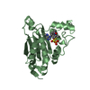
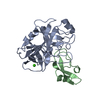
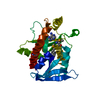
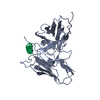
- PDB-1yk9: Crystal structure of a mutant form of the mycobacterial adenylyl ... -
+ Open data

- Basic information
| Entry | Database: PDB / ID: 1yk9 | ||||||
|---|---|---|---|---|---|---|---|
| Title | Crystal structure of a mutant form of the mycobacterial adenylyl cyclase Rv1625c | ||||||
 Components Components | Adenylate cyclase Adenylyl cyclase Adenylyl cyclase | ||||||
 Keywords Keywords | LYASE / beta-alpha-beta sandwich / Structural Genomics / PSI / Protein Structure Initiative / TB Structural Genomics Consortium / TBSGC | ||||||
| Function / homology |  Function and homology information Function and homology informationreceptor guanylyl cyclase signaling pathway / peptide receptor activity / cGMP biosynthetic process / guanylate cyclase activity / adenylate cyclase / cAMP biosynthetic process / adenylate cyclase activity / manganese ion binding / membrane => GO:0016020 / intracellular signal transduction ...receptor guanylyl cyclase signaling pathway / peptide receptor activity / cGMP biosynthetic process / guanylate cyclase activity / adenylate cyclase / cAMP biosynthetic process / adenylate cyclase activity / manganese ion binding / membrane => GO:0016020 / intracellular signal transduction / magnesium ion binding / signal transduction / ATP binding / plasma membraneSimilarity search - Function | ||||||
| Biological species |   Mycobacterium tuberculosis (bacteria) Mycobacterium tuberculosis (bacteria) | ||||||
| Method | X-RAY DIFFRACTION / SYNCHROTRON / MOLECULAR REPLACEMENT / Resolution: 2.7 Å | ||||||
 Authors Authors | Ketkar, A.D. / Shenoy, A.R. / Ramagopal, U.A. / Visweswariah, S.S. / Suguna, K. / TB Structural Genomics Consortium (TBSGC) | ||||||
 Citation Citation | Journal: J.Mol.Biol. / Year: 2006 Title: A Structural Basis for the Role of Nucleotide Specifying Residues in Regulating the Oligomerization of the Rv1625c Adenylyl Cyclase from M.tuberculosis Authors: Ketkar, A.D. / Shenoy, A.R. / Ramagopal, U.A. / Visweswariah, S.S. / Suguna, K. | ||||||
| History |
|
- Structure visualization
| Structure viewer | Molecule: MolmilJmol/JSmol |
|---|
- Downloads & links
-Download
| PDBx/mmCIF format | 1yk9.cif.gz | 50.2 KB | Display | PDBx/mmCIF format |
|---|---|---|---|---|
| PDB format | pdb1yk9.ent.gz | 35.4 KB | Display | PDB format |
| PDBx/mmJSON format | 1yk9.json.gz | Tree view | PDBx/mmJSON format | |
| Others |  Other downloads Other downloads |
-Validation report
| Arichive directory | https://data.pdbj.org/pub/pdb/validation_reports/yk/1yk9ftp://data.pdbj.org/pub/pdb/validation_reports/yk/1yk9 | HTTPS FTP |
|---|
-Related structure data
| Similar structure data |
|---|


-Links
 PDBj
PDBj


- Assembly
| Deposited unit | 
| ||||||||
|---|---|---|---|---|---|---|---|---|---|
| 1 |
| ||||||||
| Unit cell |
|
-Components
| #1: Protein | Adenylyl cyclase / ATP pyrophosphate-lyase / Adenylyl cyclase Mass: 22372.295 Da / Num. of mol.: 1 / Fragment: Rv1625c catalytic domain / Mutation: K296E, F363R, D365C Source method: isolated from a genetically manipulated source Source: (gene. exp.) Mycobacterium tuberculosis (bacteria) / Gene: Rv1625c / Plasmid: pRSET / Species (production host): Escherichia coli / Production host: Escherichia coli BL21 (bacteria) / Strain (production host): BL21References: UniProt: O30820, UniProt: P9WQ35*PLUS, adenylate cyclase |
|---|---|
| #2: Water | ChemComp-HOH / Water Mass: 18.015 Da / Num. of mol.: 71 / Source method: isolated from a natural source / Formula: H2O Mass: 18.015 Da / Num. of mol.: 71 / Source method: isolated from a natural source / Formula: H2O |
-Experimental details
-Experiment
| Experiment | Method: X-RAY DIFFRACTION / Number of used crystals: 1 |
|---|
- Sample preparation
| Crystal | Density Matthews: 2.66 Å3/Da / Density % sol: 53.4 % |
|---|---|
| Crystal grow | Temperature: 293 K / Method: vapor diffusion, hanging drop / pH: 8.5 Details: PEG 8000, Tris-HCl, pH 8.5, VAPOR DIFFUSION, HANGING DROP, temperature 293K |
-Data collection
| Diffraction | Mean temperature: 100 K |
|---|---|
| Diffraction source | Source: SYNCHROTRON / Site: NSLS  / Beamline: X9A / Wavelength: 0.9793 Å / Beamline: X9A / Wavelength: 0.9793 Å |
| Detector | Type: MARRESEARCH / Detector: CCD / Date: Jun 1, 2004 |
| Radiation | Monochromator: sagitally focusing Si(111) crystals / Protocol: SINGLE WAVELENGTH / Monochromatic (M) / Laue (L): M / Scattering type: x-ray |
| Radiation wavelength | Wavelength: 0.9793 Å / Relative weight: 1 |
| Reflection | Resolution: 2.65→19.98 Å / Num. all: 6356 / Num. obs: 6355 / % possible obs: 99.3 % / Observed criterion σ(F): 2 / Observed criterion σ(I): 2 / Redundancy: 4 % / Biso Wilson estimate: 9.5 Å2 / Rmerge(I) obs: 0.109 / Net I/σ(I): 11.8 |
| Reflection shell | Resolution: 2.65→2.74 Å / Redundancy: 3.9 % / Rmerge(I) obs: 0.46 / Mean I/σ(I) obs: 2.8 / Num. unique all: 628 / % possible all: 100 |
- Processing
| Software |
| ||||||||||||||||||||||||||||||||||||
|---|---|---|---|---|---|---|---|---|---|---|---|---|---|---|---|---|---|---|---|---|---|---|---|---|---|---|---|---|---|---|---|---|---|---|---|---|---|
| Refinement | Method to determine structure: MOLECULAR REPLACEMENT / Resolution: 2.7→19.98 Å / Rfactor Rfree error: 0.01 / Data cutoff high absF: 2007090.86 / Data cutoff low absF: 0 / Isotropic thermal model: RESTRAINED / Cross valid method: THROUGHOUT / Stereochemistry target values: Engh & Huber
| ||||||||||||||||||||||||||||||||||||
| Solvent computation | Solvent model: FLAT MODEL / Bsol: 300 Å2 / ksol: 1.16565 e/Å3 | ||||||||||||||||||||||||||||||||||||
| Displacement parameters | Biso mean: 34.5 Å2
| ||||||||||||||||||||||||||||||||||||
| Refine analyze |
| ||||||||||||||||||||||||||||||||||||
| Refinement step | Cycle: LAST / Resolution: 2.7→19.98 Å
| ||||||||||||||||||||||||||||||||||||
| Refine LS restraints |
| ||||||||||||||||||||||||||||||||||||
| LS refinement shell | Resolution: 2.7→2.87 Å / Rfactor Rfree error: 0.039 / Total num. of bins used: 6
| ||||||||||||||||||||||||||||||||||||
| Xplor file |
|
