 Movie
Movie Controller

[English] 日本語
 Yorodumi
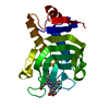
- PDB-3gqq: Crystal structure of the human retinal protein 4 (unc-119 homolog... -
+ Open data

- Basic information
| Entry | Database: PDB / ID: 3gqq | ||||||
|---|---|---|---|---|---|---|---|
| Title | Crystal structure of the human retinal protein 4 (unc-119 homolog A). Northeast Structural Genomics Consortium target HR3066a | ||||||
 Components Components | Protein unc-119 homolog A | ||||||
 Keywords Keywords |  SPLICING / Human retinal protein 4 / unc-119 homolog A / hRG4 / U119A_HUMAN / HR3066a / NESG / Structural Genomics / PSI-2 / Protein Structure Initiative / Northeast Structural Genomics Consortium / Alternative splicing / Phosphoprotein / Sensory transduction / Vision SPLICING / Human retinal protein 4 / unc-119 homolog A / hRG4 / U119A_HUMAN / HR3066a / NESG / Structural Genomics / PSI-2 / Protein Structure Initiative / Northeast Structural Genomics Consortium / Alternative splicing / Phosphoprotein / Sensory transduction / Vision | ||||||
| Function / homology |  Function and homology information Function and homology informationnegative regulation of caveolin-mediated endocytosis / negative regulation of clathrin-dependent endocytosis / lipoprotein transport / intercellular bridge / phototransduction / mitotic cytokinesis / positive regulation of protein tyrosine kinase activity / spindle midzone / visual perception / spindle pole ...negative regulation of caveolin-mediated endocytosis / negative regulation of clathrin-dependent endocytosis / lipoprotein transport / intercellular bridge / phototransduction / mitotic cytokinesis / positive regulation of protein tyrosine kinase activity / spindle midzone / visual perception / spindle pole / endocytosis / nervous system development / chemical synaptic transmission / centrosome / lipid binding / synapse / cytosolSimilarity search - Function | ||||||
| Biological species |  Homo sapiens (human) Homo sapiens (human) | ||||||
| Method | X-RAY DIFFRACTION / SYNCHROTRON / SAD / Resolution: 1.945 Å | ||||||
 Authors Authors | Vorobiev, S.M. / Chen, Y. / Seetharaman, J. / Shastry, R. / Foote, E.L. / Ciccosanti, C. / Sahdev, S. / Xiao, R. / Acton, T.B. / Montelione, G.T. ...Vorobiev, S.M. / Chen, Y. / Seetharaman, J. / Shastry, R. / Foote, E.L. / Ciccosanti, C. / Sahdev, S. / Xiao, R. / Acton, T.B. / Montelione, G.T. / Hunt, J.F. / Tong, L. / Northeast Structural Genomics Consortium (NESG) | ||||||
 Citation Citation | Journal: To be Published Title: Crystal structure of the human retinal protein 4 (unc-119 homolog A). Authors: Vorobiev, S.M. / Chen, Y. / Seetharaman, J. / Shastry, R. / Foote, E.L. / Ciccosanti, C. / Sahdev, S. / Xiao, R. / Acton, T.B. / Montelione, G.T. / Hunt, J.F. / Tong, L. | ||||||
| History |
|
- Structure visualization
| Structure viewer | Molecule: MolmilJmol/JSmol |
|---|
- Downloads & links
-Download
| PDBx/mmCIF format | 3gqq.cif.gz | 439.4 KB | Display | PDBx/mmCIF format |
|---|---|---|---|---|
| PDB format | pdb3gqq.ent.gz | 375.3 KB | Display | PDB format |
| PDBx/mmJSON format | 3gqq.json.gz | Tree view | PDBx/mmJSON format | |
| Others |  Other downloads Other downloads |
-Validation report
| Arichive directory | https://data.pdbj.org/pub/pdb/validation_reports/gq/3gqqftp://data.pdbj.org/pub/pdb/validation_reports/gq/3gqq | HTTPS FTP |
|---|
-Related structure data
| Similar structure data | |
|---|---|
| Other databases |


-Links
 PDBj
PDBj

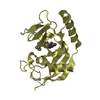
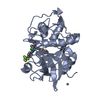
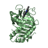
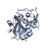
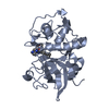
- Assembly
| Deposited unit | 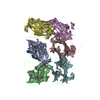
| ||||||||
|---|---|---|---|---|---|---|---|---|---|
| 1 | 
| ||||||||
| 2 | 
| ||||||||
| 3 | 
| ||||||||
| 4 | 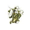
| ||||||||
| 5 | 
| ||||||||
| 6 | 
| ||||||||
| Unit cell |
| ||||||||
| Details | Dimer according to aggregation screening |
-Components
| #1: Protein | Mass: 23293.604 Da / Num. of mol.: 6 / Fragment: UNP residues 56-240 Source method: isolated from a genetically manipulated source Source: (gene. exp.) Homo sapiens (human) / Gene: RG4, UNC119 / Plasmid: pET14-15C / Production host:  Escherichia coli (E. coli) / Strain (production host): BL21(DE3)+Magic / References: UniProt: Q13432 Escherichia coli (E. coli) / Strain (production host): BL21(DE3)+Magic / References: UniProt: Q13432#2: Chemical | ChemComp-UNL / Num. of mol.: 30 / Source method: obtained synthetically #3: Water | ChemComp-HOH / | Water Mass: 18.015 Da / Num. of mol.: 803 / Source method: isolated from a natural source / Formula: H2O Mass: 18.015 Da / Num. of mol.: 803 / Source method: isolated from a natural source / Formula: H2O |
|---|
-Experimental details
-Experiment
| Experiment | Method: X-RAY DIFFRACTION / Number of used crystals: 1 |
|---|
- Sample preparation
| Crystal | Density Matthews: 2.1 Å3/Da / Density % sol: 41.51 % Description: The structure factor file contains Friedel pairs |
|---|---|
| Crystal grow | Temperature: 277 K / Method: microbatch under oil / pH: 5 Details: 40% PEG 4000, 0.1M K acetate, 0.1M Na acetate, pH 5.0, MICROBATCH UNDER OIL, temperature 277K |
-Data collection
| Diffraction | Mean temperature: 100 K |
|---|---|
| Diffraction source | Source: SYNCHROTRON / Site: NSLS  / Beamline: X4A / Wavelength: 0.97934 Å / Beamline: X4A / Wavelength: 0.97934 Å |
| Detector | Type: ADSC QUANTUM 4 / Detector: CCD / Date: Feb 23, 2009 |
| Radiation | Protocol: SINGLE WAVELENGTH / Monochromatic (M) / Laue (L): M / Scattering type: x-ray |
| Radiation wavelength | Wavelength: 0.97934 Å / Relative weight: 1 |
| Reflection | Resolution: 1.945→50 Å / Num. all: 166290 / Num. obs: 166290 / % possible obs: 99.6 % / Observed criterion σ(F): 0 / Observed criterion σ(I): 0 / Redundancy: 7 % / Rmerge(I) obs: 0.088 / Net I/σ(I): 26.4 |
| Reflection shell | Resolution: 1.945→2.02 Å / Redundancy: 4.8 % / Rmerge(I) obs: 0.537 / Mean I/σ(I) obs: 2.87 / Num. unique all: 16637 / % possible all: 100 |
- Processing
| Software |
| ||||||||||||||||||||||||||||||||||||||||||||||||||||||||||||||||||||||||||||||||||||||||||||||||||||||||||||||||||||||||||||||||||||||||||||||||||||||||||||||||||||||||||||||||||||||||||
|---|---|---|---|---|---|---|---|---|---|---|---|---|---|---|---|---|---|---|---|---|---|---|---|---|---|---|---|---|---|---|---|---|---|---|---|---|---|---|---|---|---|---|---|---|---|---|---|---|---|---|---|---|---|---|---|---|---|---|---|---|---|---|---|---|---|---|---|---|---|---|---|---|---|---|---|---|---|---|---|---|---|---|---|---|---|---|---|---|---|---|---|---|---|---|---|---|---|---|---|---|---|---|---|---|---|---|---|---|---|---|---|---|---|---|---|---|---|---|---|---|---|---|---|---|---|---|---|---|---|---|---|---|---|---|---|---|---|---|---|---|---|---|---|---|---|---|---|---|---|---|---|---|---|---|---|---|---|---|---|---|---|---|---|---|---|---|---|---|---|---|---|---|---|---|---|---|---|---|---|---|---|---|---|---|---|---|---|
| Refinement | Method to determine structure: SAD / Resolution: 1.945→40.51 Å / SU ML: 0.34 / Cross valid method: THROUGHOUT / Stereochemistry target values: MAXIMUM LIKELIHOOD Details: The Friedel pairs were used in phasing. An unknown ligand bound to each hRG4 molecules has been refined. The unknown ligand (UNL) has been modeled in electron density by water molecules.
| ||||||||||||||||||||||||||||||||||||||||||||||||||||||||||||||||||||||||||||||||||||||||||||||||||||||||||||||||||||||||||||||||||||||||||||||||||||||||||||||||||||||||||||||||||||||||||
| Solvent computation | Shrinkage radii: 0.9 Å / VDW probe radii: 1.11 Å / Solvent model: FLAT BULK SOLVENT MODEL / Bsol: 53.318 Å2 / ksol: 0.343 e/Å3 | ||||||||||||||||||||||||||||||||||||||||||||||||||||||||||||||||||||||||||||||||||||||||||||||||||||||||||||||||||||||||||||||||||||||||||||||||||||||||||||||||||||||||||||||||||||||||||
| Refinement step | Cycle: LAST / Resolution: 1.945→40.51 Å
| ||||||||||||||||||||||||||||||||||||||||||||||||||||||||||||||||||||||||||||||||||||||||||||||||||||||||||||||||||||||||||||||||||||||||||||||||||||||||||||||||||||||||||||||||||||||||||
| Refine LS restraints |
| ||||||||||||||||||||||||||||||||||||||||||||||||||||||||||||||||||||||||||||||||||||||||||||||||||||||||||||||||||||||||||||||||||||||||||||||||||||||||||||||||||||||||||||||||||||||||||
| LS refinement shell | Refine-ID: X-RAY DIFFRACTION / Total num. of bins used: 30
| ||||||||||||||||||||||||||||||||||||||||||||||||||||||||||||||||||||||||||||||||||||||||||||||||||||||||||||||||||||||||||||||||||||||||||||||||||||||||||||||||||||||||||||||||||||||||||
| Refinement TLS params. | Method: refined / Refine-ID: X-RAY DIFFRACTION
| ||||||||||||||||||||||||||||||||||||||||||||||||||||||||||||||||||||||||||||||||||||||||||||||||||||||||||||||||||||||||||||||||||||||||||||||||||||||||||||||||||||||||||||||||||||||||||
| Refinement TLS group |
|
