 Movie
Movie Controller


+ Open data

- Basic information
| Entry | Database: PDB / ID: 1yht | ||||||
|---|---|---|---|---|---|---|---|
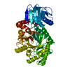
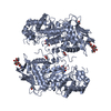
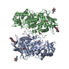
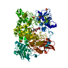
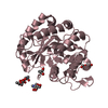
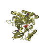
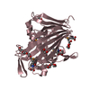
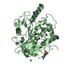
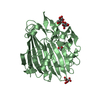
| Title | Crystal structure analysis of Dispersin B | ||||||
 Components Components | DspB | ||||||
 Keywords Keywords |  HYDROLASE / beta barrel HYDROLASE / beta barrel | ||||||
| Function / homology |  Function and homology informationbeta-N-acetylhexosaminidase activity / beta-N-acetylhexosaminidase / N-acetyl-beta-D-galactosaminidase activity / carbohydrate metabolic process Function and homology informationbeta-N-acetylhexosaminidase activity / beta-N-acetylhexosaminidase / N-acetyl-beta-D-galactosaminidase activity / carbohydrate metabolic processSimilarity search - Function | ||||||
| Biological species |  Aggregatibacter actinomycetemcomitans (bacteria) Aggregatibacter actinomycetemcomitans (bacteria) | ||||||
| Method | X-RAY DIFFRACTION / SYNCHROTRON / MAD / Resolution: 2 Å | ||||||
 Authors Authors | Ramasubbu, N. / Thomas, L.M. / Ragunath, C. / Kaplan, J.B. | ||||||
 Citation Citation | Journal: J.Mol.Biol. / Year: 2005 Title: Structural Analysis of Dispersin B, a Biofilm-releasing Glycoside Hydrolase from the Periodontopathogen Actinobacillus actinomycetemcomitans. Authors: Ramasubbu, N. / Thomas, L.M. / Ragunath, C. / Kaplan, J.B. #1: Journal: J.Bacteriol. / Year: 2003 Title: Detachment of Actinobacillus actinomycetemcomitans biofilm cells by an endogenous beta-hexosaminidase activity Authors: Kaplan, J.B. / Ragunath, C. / Ramasubbu, N. / Fine, D.H. #2: Journal: Antimicrob.Agents Chemother. / Year: 2004 Title: Enzymatic detachment of Staphylococcus epidermidis biofilms Authors: Kaplan, J.B. / Ragunath, C. / Velliyagounder, K. / Fine, D.H. / Ramasubbu, N. #3: Journal: J.Bacteriol. / Year: 2004 Title: Genes involved in the synthesis and degradation of matrix polysaccharide in Actinobacillus actinomycetemcomitans and Actinobacillus pleuropneumoniae biofilms. Authors: Kaplan, J.B. / Velliyagounder, K. / Ragunath, C. / Rhode, H. / Mack, D. / Knobloch, J.K. / Ramasubbu, N. | ||||||
| History |
|
- Structure visualization
| Structure viewer | Molecule: MolmilJmol/JSmol |
|---|
- Downloads & links
-Download
| PDBx/mmCIF format | 1yht.cif.gz | 87 KB | Display | PDBx/mmCIF format |
|---|---|---|---|---|
| PDB format | pdb1yht.ent.gz | 65.5 KB | Display | PDB format |
| PDBx/mmJSON format | 1yht.json.gz | Tree view | PDBx/mmJSON format | |
| Others |  Other downloads Other downloads |
-Validation report
| Arichive directory | https://data.pdbj.org/pub/pdb/validation_reports/yh/1yhtftp://data.pdbj.org/pub/pdb/validation_reports/yh/1yht | HTTPS FTP |
|---|
-Related structure data
| Related structure data | |
|---|---|
| Similar structure data |


-Links
 PDBj
PDBj

- Assembly
| Deposited unit | 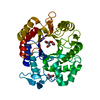
| |||||||||
|---|---|---|---|---|---|---|---|---|---|---|
| 1 |
| |||||||||
| Unit cell |
| |||||||||
| Components on special symmetry positions |
|
-Components
| #1: Protein | Mass: 41842.797 Da / Num. of mol.: 1 Source method: isolated from a genetically manipulated source Source: (gene. exp.) Aggregatibacter actinomycetemcomitans (bacteria)Strain: CU1000 / Gene: dspB Plasmid details: The dspB gene was inserted into pET29B with 6 His tags at the C-terminus leading to the plasmid pRC3. Plasmid: pET29b / Production host: Escherichia coli (E. coli)References: GenBank: 30420960, UniProt: Q840G9*PLUS, beta-N-acetylhexosaminidase | ||
|---|---|---|---|
| #2: Chemical | ChemComp-ACY / Acetic acid  Mass: 60.052 Da / Num. of mol.: 1 / Source method: obtained synthetically / Formula: C2H4O2 Mass: 60.052 Da / Num. of mol.: 1 / Source method: obtained synthetically / Formula: C2H4O2 | ||
| #3: Chemical | Glycerol  Mass: 92.094 Da / Num. of mol.: 2 / Source method: obtained synthetically / Formula: C3H8O3 Mass: 92.094 Da / Num. of mol.: 2 / Source method: obtained synthetically / Formula: C3H8O3#4: Water | ChemComp-HOH / | Water Mass: 18.015 Da / Num. of mol.: 243 / Source method: isolated from a natural source / Formula: H2O Mass: 18.015 Da / Num. of mol.: 243 / Source method: isolated from a natural source / Formula: H2O |
-Experimental details
-Experiment
| Experiment | Method: X-RAY DIFFRACTION / Number of used crystals: 1 |
|---|
- Sample preparation
| Crystal | Density Matthews: 2 Å3/Da / Density % sol: 38 % |
|---|---|
| Crystal grow | Temperature: 298 K / Method: vapor diffusion, hanging drop / pH: 5.5 Details: PEG 10000, ammonium acetate, BisTris, pH 5.5, VAPOR DIFFUSION, HANGING DROP, temperature 298K |
-Data collection
| Diffraction |
| ||||||||||||||||||
|---|---|---|---|---|---|---|---|---|---|---|---|---|---|---|---|---|---|---|---|
| Diffraction source | Source: SYNCHROTRON / Site: SSRL  / Beamline: BL9-2 / Wavelength: 0.91837, 0.979 / Beamline: BL9-2 / Wavelength: 0.91837, 0.979 | ||||||||||||||||||
| Detector | Type: ADSC QUANTUM 315 / Detector: CCD / Date: Jun 1, 2004 | ||||||||||||||||||
| Radiation |
| ||||||||||||||||||
| Radiation wavelength |
| ||||||||||||||||||
| Reflection | Resolution: 2→23.76 Å / Num. all: 22562 / Num. obs: 21412 / % possible obs: 99.1 % / Observed criterion σ(F): 2 / Observed criterion σ(I): 2 / Redundancy: 4.1 % / Rmerge(I) obs: 0.071 / Net I/σ(I): 15.6 | ||||||||||||||||||
| Reflection shell | Resolution: 2→2.1 Å / Redundancy: 4 % / Rmerge(I) obs: 0.193 / Mean I/σ(I) obs: 7.1 / % possible all: 99.9 |
- Processing
| Software |
| |||||||||||||||||||||||||||||||||||||||||||||||||||||||||||||||||||||||||||||||||||||||||||||||||||||||||||||||||||||||||||||
|---|---|---|---|---|---|---|---|---|---|---|---|---|---|---|---|---|---|---|---|---|---|---|---|---|---|---|---|---|---|---|---|---|---|---|---|---|---|---|---|---|---|---|---|---|---|---|---|---|---|---|---|---|---|---|---|---|---|---|---|---|---|---|---|---|---|---|---|---|---|---|---|---|---|---|---|---|---|---|---|---|---|---|---|---|---|---|---|---|---|---|---|---|---|---|---|---|---|---|---|---|---|---|---|---|---|---|---|---|---|---|---|---|---|---|---|---|---|---|---|---|---|---|---|---|---|---|
| Refinement | Method to determine structure: MAD / Resolution: 2→23.76 Å / Cor.coef. Fo:Fc: 0.955 / Cor.coef. Fo:Fc free: 0.916 / SU B: 3.438 / SU ML: 0.099 / Cross valid method: THROUGHOUT / σ(F): 0 / ESU R: 0.176 / ESU R Free: 0.162 / Stereochemistry target values: MAXIMUM LIKELIHOOD Details: HYDROGENS HAVE BEEN ADDED IN THE RIDING POSITIONS. The electron density for residues Tyr187, Ser188, Val189, Glu190 and Ser191 are not well resolved.
| |||||||||||||||||||||||||||||||||||||||||||||||||||||||||||||||||||||||||||||||||||||||||||||||||||||||||||||||||||||||||||||
| Solvent computation | Ion probe radii: 0.8 Å / Shrinkage radii: 0.8 Å / VDW probe radii: 1.2 Å / Solvent model: BABINET MODEL WITH MASK | |||||||||||||||||||||||||||||||||||||||||||||||||||||||||||||||||||||||||||||||||||||||||||||||||||||||||||||||||||||||||||||
| Displacement parameters | Biso mean: 12.755 Å2
| |||||||||||||||||||||||||||||||||||||||||||||||||||||||||||||||||||||||||||||||||||||||||||||||||||||||||||||||||||||||||||||
| Refinement step | Cycle: LAST / Resolution: 2→23.76 Å
| |||||||||||||||||||||||||||||||||||||||||||||||||||||||||||||||||||||||||||||||||||||||||||||||||||||||||||||||||||||||||||||
| Refine LS restraints |
| |||||||||||||||||||||||||||||||||||||||||||||||||||||||||||||||||||||||||||||||||||||||||||||||||||||||||||||||||||||||||||||
| LS refinement shell | Resolution: 2→2.053 Å / Total num. of bins used: 20
|
