 Movie
Movie Controller


+ Open data

- Basic information
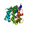
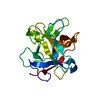
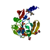
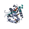
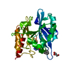
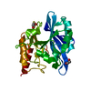
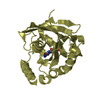
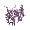
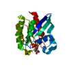
| Entry | Database: PDB / ID: 1ygh | ||||||
|---|---|---|---|---|---|---|---|
| Title | HAT DOMAIN OF GCN5 FROM SACCHAROMYCES CEREVISIAE | ||||||
 Components Components | PROTEIN (TRANSCRIPTIONAL ACTIVATOR GCN5) | ||||||
 Keywords Keywords |  GENE REGULATION / TRANSCRIPTIONAL REGULATION / HISTONE ACETYLATION / N-ACETYLTRANSFERASE / GCN5 RELATED N-ACETYLTRANSFERASE FAMILY GENE REGULATION / TRANSCRIPTIONAL REGULATION / HISTONE ACETYLATION / N-ACETYLTRANSFERASE / GCN5 RELATED N-ACETYLTRANSFERASE FAMILY | ||||||
| Function / homology |  Function and homology information Function and homology informationADA complex / histone crotonyltransferase activity / histone H3 acetyltransferase activity / SLIK (SAGA-like) complex / SAGA complex / peptide-lysine-N-acetyltransferase activity / Estrogen-dependent gene expression / chromosome, centromeric region / histone acetyltransferase complex / histone acetyltransferase activity ...ADA complex / histone crotonyltransferase activity / histone H3 acetyltransferase activity / SLIK (SAGA-like) complex / SAGA complex / peptide-lysine-N-acetyltransferase activity / Estrogen-dependent gene expression / chromosome, centromeric region / histone acetyltransferase complex / histone acetyltransferase activity / Transferases; Acyltransferases; Transferring groups other than aminoacyl groups / histone acetyltransferase / positive regulation of transcription elongation by RNA polymerase II / transcription coregulator activity / lysine-acetylated histone binding / chromatin organization / chromatin remodeling / regulation of transcription by RNA polymerase II / positive regulation of transcription by RNA polymerase II / nucleus / cytosolSimilarity search - Function | ||||||
| Biological species |  Saccharomyces cerevisiae (brewer's yeast) Saccharomyces cerevisiae (brewer's yeast) | ||||||
| Method | X-RAY DIFFRACTION / MOLECULAR REPLACEMENT / Resolution: 1.9 Å | ||||||
 Authors Authors | Trievel, R.C. / Rojas, J.R. / Sterner, D.E. / Venkataramani, R. / Wang, L. / Zhou, J. / Allis, C.D. / Berger, S.L. / Marmorstein, R. | ||||||
 Citation Citation | Journal: Proc.Natl.Acad.Sci.USA / Year: 1999 Title: Crystal structure and mechanism of histone acetylation of the yeast GCN5 transcriptional coactivator. Authors: Trievel, R.C. / Rojas, J.R. / Sterner, D.E. / Venkataramani, R.N. / Wang, L. / Zhou, J. / Allis, C.D. / Berger, S.L. / Marmorstein, R. | ||||||
| History |
|
- Structure visualization
| Structure viewer | Molecule: MolmilJmol/JSmol |
|---|
- Downloads & links
-Download
| PDBx/mmCIF format | 1ygh.cif.gz | 82.1 KB | Display | PDBx/mmCIF format |
|---|---|---|---|---|
| PDB format | pdb1ygh.ent.gz | 62.1 KB | Display | PDB format |
| PDBx/mmJSON format | 1ygh.json.gz | Tree view | PDBx/mmJSON format | |
| Others |  Other downloads Other downloads |
-Validation report
| Arichive directory | https://data.pdbj.org/pub/pdb/validation_reports/yg/1yghftp://data.pdbj.org/pub/pdb/validation_reports/yg/1ygh | HTTPS FTP |
|---|
-Related structure data
| Similar structure data |
|---|

-Links
 PDBj
PDBj


- Assembly
| Deposited unit | 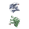
| ||||||||
|---|---|---|---|---|---|---|---|---|---|
| 1 | 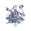
| ||||||||
| 2 | 
| ||||||||
| Unit cell |
| ||||||||
| Noncrystallographic symmetry (NCS) | NCS oper: (Code: given Matrix: (0.999966, 0.005455, 0.0062), Vector : |
-Components
| #1: Protein | Mass: 19248.455 Da / Num. of mol.: 2 / Fragment: HISTONE ACETYLTRANSFERASE DOMAIN Source method: isolated from a genetically manipulated source Source: (gene. exp.) Saccharomyces cerevisiae (brewer's yeast)Cellular location: NUCLEAR / Gene: GCN5 / Plasmid: PRSET / Species (production host): Escherichia coli / Gene (production host): GCN5 / Production host:  Escherichia coli BL21(DE3) (bacteria) / Strain (production host): BL21(DE3) / References: UniProt: Q03330, histone acetyltransferase Escherichia coli BL21(DE3) (bacteria) / Strain (production host): BL21(DE3) / References: UniProt: Q03330, histone acetyltransferase#2: Chemical | Glycerol  Mass: 92.094 Da / Num. of mol.: 2 / Source method: obtained synthetically / Formula: C3H8O3 Mass: 92.094 Da / Num. of mol.: 2 / Source method: obtained synthetically / Formula: C3H8O3#3: Water | ChemComp-HOH / | Water Mass: 18.015 Da / Num. of mol.: 216 / Source method: isolated from a natural source / Formula: H2O Mass: 18.015 Da / Num. of mol.: 216 / Source method: isolated from a natural source / Formula: H2O |
|---|
-Experimental details
-Experiment
| Experiment | Method: X-RAY DIFFRACTION / Number of used crystals: 1 |
|---|
- Sample preparation
| Crystal | Density Matthews: 2.7 Å3/Da / Density % sol: 55 % | ||||||||||||||||||||
|---|---|---|---|---|---|---|---|---|---|---|---|---|---|---|---|---|---|---|---|---|---|
| Crystal grow | pH: 6 Details: 400 MM AMMONIUM SULFATE, 20% - 25% PEG 8000, pH 6.0 Temp details: 277 | ||||||||||||||||||||
| Crystal | *PLUS | ||||||||||||||||||||
| Crystal grow | *PLUS Temperature: 4 ℃ / Method: vapor diffusion, hanging drop | ||||||||||||||||||||
| Components of the solutions | *PLUS
|
-Data collection
| Diffraction | Mean temperature: 93 K |
|---|---|
| Diffraction source | Source: ROTATING ANODE / Type: RIGAKU RU300 / Wavelength: 1.5418 |
| Detector | Type: RIGAKU RAXIS IV / Detector: IMAGE PLATE / Date: Oct 1, 1998 / Details: MIRRORS |
| Radiation | Monochromator: NI FILTER / Protocol: SINGLE WAVELENGTH / Monochromatic (M) / Laue (L): M / Scattering type: x-ray |
| Radiation wavelength | Wavelength: 1.5418 Å / Relative weight: 1 |
| Reflection | Resolution: 1.9→50 Å / Num. obs: 31832 / % possible obs: 96.4 % / Observed criterion σ(I): -3 / Redundancy: 5.7 % / Biso Wilson estimate: 18.6 Å2 / Rsym value: 0.03 / Net I/σ(I): 23.6 |
| Reflection shell | Resolution: 1.9→1.94 Å / Mean I/σ(I) obs: 13 / Rsym value: 0.088 / % possible all: 98 |
| Reflection | *PLUS Num. measured all: 181066 / Rmerge(I) obs: 0.03 |
- Processing
| Software |
| ||||||||||||||||||||||||||||||||||||||||||||||||||||||||||||
|---|---|---|---|---|---|---|---|---|---|---|---|---|---|---|---|---|---|---|---|---|---|---|---|---|---|---|---|---|---|---|---|---|---|---|---|---|---|---|---|---|---|---|---|---|---|---|---|---|---|---|---|---|---|---|---|---|---|---|---|---|---|
| Refinement | Method to determine structure: MOLECULAR REPLACEMENT Starting model: TETRAHYMENA THERMOPHILA GCN5 HAT DOMAIN Resolution: 1.9→50 Å / Cross valid method: THROUGHOUT / σ(F): 2
| ||||||||||||||||||||||||||||||||||||||||||||||||||||||||||||
| Displacement parameters | Biso mean: 16.9 Å2 | ||||||||||||||||||||||||||||||||||||||||||||||||||||||||||||
| Refinement step | Cycle: LAST / Resolution: 1.9→50 Å
| ||||||||||||||||||||||||||||||||||||||||||||||||||||||||||||
| Refine LS restraints |
| ||||||||||||||||||||||||||||||||||||||||||||||||||||||||||||
| LS refinement shell | Resolution: 1.9→1.99 Å / Total num. of bins used: 8
| ||||||||||||||||||||||||||||||||||||||||||||||||||||||||||||
| Xplor file |
| ||||||||||||||||||||||||||||||||||||||||||||||||||||||||||||
| Software | *PLUS Name: X-PLOR / Version: 3.851 / Classification: refinement | ||||||||||||||||||||||||||||||||||||||||||||||||||||||||||||
| Refinement | *PLUS σ(F): 2 / % reflection Rfree: 10 % | ||||||||||||||||||||||||||||||||||||||||||||||||||||||||||||
| Solvent computation | *PLUS | ||||||||||||||||||||||||||||||||||||||||||||||||||||||||||||
| Displacement parameters | *PLUS Biso mean: 16.9 Å2 | ||||||||||||||||||||||||||||||||||||||||||||||||||||||||||||
| LS refinement shell | *PLUS Rfactor Rfree: 0.269 / Rfactor Rwork: 0.261 |
