 Movie
Movie Controller


+ Open data

- Basic information
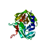
| Entry | Database: PDB / ID: 4h9w | ||||||
|---|---|---|---|---|---|---|---|
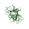
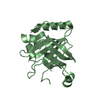
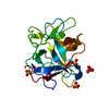
| Title | crystal structure of a METHIONINE mutant of WCI | ||||||
 Components Components | Chymotrypsin inhibitor 3 | ||||||
 Keywords Keywords | HYDROLASE INHIBITOR / chymotrypsin inhibitor / inhibitor of chymotrypsin | ||||||
| Function / homology |  Function and homology information Function and homology information | ||||||
| Biological species |    Psophocarpus tetragonolobus (winged bean) Psophocarpus tetragonolobus (winged bean) | ||||||
| Method | X-RAY DIFFRACTION / MOLECULAR REPLACEMENT / Resolution: 2.5 Å | ||||||
 Authors Authors | Majumder, S. / Sen, U. | ||||||
 Citation Citation | Journal: Biochim.Biophys.Acta / Year: 2015 Title: A conserved tryptophan (W91) at the barrel-lid junction modulates the packing and stability of Kunitz (STI) family of inhibitors. Authors: Majumder, S. / Khamrui, S. / Banerjee, R. / Bhowmik, P. / Sen, U. | ||||||
| History |
|
- Structure visualization
| Structure viewer | Molecule: MolmilJmol/JSmol |
|---|
- Downloads & links
-Download
| PDBx/mmCIF format | 4h9w.cif.gz | 50.5 KB | Display | PDBx/mmCIF format |
|---|---|---|---|---|
| PDB format | pdb4h9w.ent.gz | 35.6 KB | Display | PDB format |
| PDBx/mmJSON format | 4h9w.json.gz | Tree view | PDBx/mmJSON format | |
| Others |  Other downloads Other downloads |
-Validation report
| Arichive directory | https://data.pdbj.org/pub/pdb/validation_reports/h9/4h9wftp://data.pdbj.org/pub/pdb/validation_reports/h9/4h9w | HTTPS FTP |
|---|
-Related structure data
| Related structure data |  4ha2C 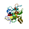 4tlpC  1eylS C: citing same article ( S: Starting model for refinement |
|---|---|
| Similar structure data |


-Links
 PDBj
PDBj- Assembly
| Deposited unit | 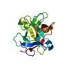
| ||||||||
|---|---|---|---|---|---|---|---|---|---|
| 1 |
| ||||||||
| Unit cell |
|
-Components
| #1: Protein | Mass: 20619.318 Da / Num. of mol.: 1 / Mutation: W91M Source method: isolated from a genetically manipulated source Source: (gene. exp.) Psophocarpus tetragonolobus (winged bean)Plasmid: PET28a / Production host:  Escherichia coli (E. coli) / Strain (production host): BL21(DE3) / References: UniProt: P10822 Escherichia coli (E. coli) / Strain (production host): BL21(DE3) / References: UniProt: P10822 |
|---|---|
| #2: Water | ChemComp-HOH / Water Mass: 18.015 Da / Num. of mol.: 142 / Source method: isolated from a natural source / Formula: H2O Mass: 18.015 Da / Num. of mol.: 142 / Source method: isolated from a natural source / Formula: H2O |
-Experimental details
-Experiment
| Experiment | Method: X-RAY DIFFRACTION / Number of used crystals: 1 |
|---|
- Sample preparation
| Crystal | Density Matthews: 2.82 Å3/Da / Density % sol: 56.32 % |
|---|---|
| Crystal grow | Temperature: 277 K / Method: vapor diffusion, hanging drop / pH: 4 Details: 0.3M AMS, 5% GLYCEROL, 25% PEG 6K, pH 4, VAPOR DIFFUSION, HANGING DROP, temperature 277K |
-Data collection
| Diffraction | Mean temperature: 100 K |
|---|---|
| Diffraction source | Source: ROTATING ANODE / Type: BRUKER AXS MICROSTAR / Wavelength: 1.5418 Å |
| Detector | Type: MAR scanner 345 mm plate / Detector: IMAGE PLATE / Date: Aug 2, 2011 |
| Radiation | Monochromator: graphaite / Protocol: SINGLE WAVELENGTH / Monochromatic (M) / Laue (L): M / Scattering type: x-ray |
| Radiation wavelength | Wavelength: 1.5418 Å / Relative weight: 1 |
| Reflection | Resolution: 2.5→30 Å / Num. all: 8943 / Num. obs: 8893 / % possible obs: 99.2 % / Observed criterion σ(F): 2 / Observed criterion σ(I): 0 / Redundancy: 7.05 % / Rmerge(I) obs: 0.055 / Net I/σ(I): 7.1 |
| Reflection shell | Highest resolution: 2.5 Å / Redundancy: 7.05 % / Rmerge(I) obs: 0.055 / Mean I/σ(I) obs: 7.1 / Num. unique all: 8943 / % possible all: 99.2 |
- Processing
| Software |
| |||||||||||||||||||||||||
|---|---|---|---|---|---|---|---|---|---|---|---|---|---|---|---|---|---|---|---|---|---|---|---|---|---|---|
| Refinement | Method to determine structure: MOLECULAR REPLACEMENT Starting model: 1EYL Resolution: 2.5→30 Å / Cross valid method: THROUGHOUT / σ(F): 1 / Stereochemistry target values: Engh & Huber
| |||||||||||||||||||||||||
| Refinement step | Cycle: LAST / Resolution: 2.5→30 Å
| |||||||||||||||||||||||||
| LS refinement shell | Highest resolution: 2.5 Å
|
