 Movie
Movie Controller


+ Open data

- Basic information
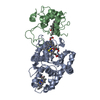
| Entry | Database: PDB / ID: 1yad | ||||||
|---|---|---|---|---|---|---|---|
| Title | Structure of TenI from Bacillus subtilis | ||||||
 Components Components | Regulatory protein tenI | ||||||
 Keywords Keywords |  TRANSCRIPTION / TIM barrel TRANSCRIPTION / TIM barrel | ||||||
| Function / homology |  Function and homology informationthiazole tautomerase / thiamine-phosphate diphosphorylase activity / thiamine diphosphate biosynthetic process / thiamine biosynthetic process / isomerase activity / cytoplasm Function and homology informationthiazole tautomerase / thiamine-phosphate diphosphorylase activity / thiamine diphosphate biosynthetic process / thiamine biosynthetic process / isomerase activity / cytoplasmSimilarity search - Function | ||||||
| Biological species |  Bacillus subtilis (bacteria) Bacillus subtilis (bacteria) | ||||||
| Method | X-RAY DIFFRACTION / SYNCHROTRON / SAD / Resolution: 2.1 Å | ||||||
 Authors Authors | Toms, A.V. / Haas, A.L. / Park, J.-H. / Begley, T.P. / Ealick, S.E. | ||||||
 Citation Citation | Journal: Biochemistry / Year: 2005 Title: Structural characterization of the regulatory proteins TenA and TenI from Bacillus subtilis and identification of TenA as a thiaminase II. Authors: Toms, A.V. / Haas, A.L. / Park, J.H. / Begley, T.P. / Ealick, S.E. | ||||||
| History |
|
- Structure visualization
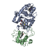
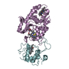
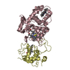
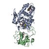
| Structure viewer | Molecule: MolmilJmol/JSmol |
|---|
- Downloads & links
-Download
| PDBx/mmCIF format | 1yad.cif.gz | 170.9 KB | Display | PDBx/mmCIF format |
|---|---|---|---|---|
| PDB format | pdb1yad.ent.gz | 135.1 KB | Display | PDB format |
| PDBx/mmJSON format | 1yad.json.gz | Tree view | PDBx/mmJSON format | |
| Others |  Other downloads Other downloads |
-Validation report
| Arichive directory | https://data.pdbj.org/pub/pdb/validation_reports/ya/1yadftp://data.pdbj.org/pub/pdb/validation_reports/ya/1yad | HTTPS FTP |
|---|
-Related structure data


-Links
 PDBj
PDBj- Assembly
| Deposited unit | 
| ||||||||
|---|---|---|---|---|---|---|---|---|---|
| 1 | 
| ||||||||
| 2 | 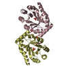
| ||||||||
| Unit cell |
| ||||||||
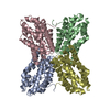
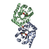
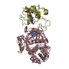
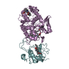
| Details | The biological assembly is a dimer. The asymmetric unit contains 2 biological units. |
-Components
| #1: Protein | Mass: 24775.539 Da / Num. of mol.: 4 Source method: isolated from a genetically manipulated source Source: (gene. exp.) Bacillus subtilis (bacteria) / Gene: tenI / Plasmid: pET-28a / Species (production host): Escherichia coli / Production host: Escherichia coli BL21(DE3) (bacteria) / Strain (production host): BL21(DE3) / References: UniProt: P25053#2: Chemical | ChemComp-SO4 / Sulfate  Mass: 96.063 Da / Num. of mol.: 8 / Source method: obtained synthetically / Formula: SO4 Mass: 96.063 Da / Num. of mol.: 8 / Source method: obtained synthetically / Formula: SO4#3: Chemical | 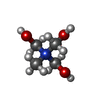  Mass: 122.143 Da / Num. of mol.: 3 / Source method: obtained synthetically / Formula: C4H12NO3 Mass: 122.143 Da / Num. of mol.: 3 / Source method: obtained synthetically / Formula: C4H12NO3#4: Water | ChemComp-HOH / | Water Mass: 18.015 Da / Num. of mol.: 543 / Source method: isolated from a natural source / Formula: H2O Mass: 18.015 Da / Num. of mol.: 543 / Source method: isolated from a natural source / Formula: H2O |
|---|
-Experimental details
-Experiment
| Experiment | Method: X-RAY DIFFRACTION / Number of used crystals: 1 |
|---|
- Sample preparation
| Crystal | Density Matthews: 2.99 Å3/Da / Density % sol: 57 % |
|---|---|
| Crystal grow | Temperature: 297 K / Method: vapor diffusion, hanging drop / pH: 9 Details: ammonium sulfate, pH 9.0, VAPOR DIFFUSION, HANGING DROP, temperature 297K |
-Data collection
| Diffraction | Mean temperature: 100 K |
|---|---|
| Diffraction source | Source: SYNCHROTRON / Site: APS  / Beamline: 8-BM / Wavelength: 0.9791 Å / Beamline: 8-BM / Wavelength: 0.9791 Å |
| Detector | Type: ADSC QUANTUM 315 / Detector: CCD / Date: Jul 24, 2003 |
| Radiation | Protocol: SINGLE WAVELENGTH / Monochromatic (M) / Laue (L): M / Scattering type: x-ray |
| Radiation wavelength | Wavelength: 0.9791 Å / Relative weight: 1 |
| Reflection | Resolution: 2.1→50 Å / Num. all: 72558 / Num. obs: 61326 / % possible obs: 95.8 % / Observed criterion σ(F): 1 / Observed criterion σ(I): 1 / Redundancy: 5.8 % / Biso Wilson estimate: 14.2 Å2 / Rsym value: 0.088 / Net I/σ(I): 18.8 |
| Reflection shell | Resolution: 2.1→2.23 Å / Redundancy: 5.8 % / Mean I/σ(I) obs: 4.3 / Num. unique all: 9652 / Rsym value: 0.396 / % possible all: 91.5 |
- Processing
| Software |
| ||||||||||||||||||||||||||||||||||||
|---|---|---|---|---|---|---|---|---|---|---|---|---|---|---|---|---|---|---|---|---|---|---|---|---|---|---|---|---|---|---|---|---|---|---|---|---|---|
| Refinement | Method to determine structure: SAD / Resolution: 2.1→37.01 Å / Rfactor Rfree error: 0.004 / Data cutoff high absF: 138911.41 / Data cutoff low absF: 0 / Isotropic thermal model: RESTRAINED / Cross valid method: THROUGHOUT / σ(F): 0 / Stereochemistry target values: Engh & Huber
| ||||||||||||||||||||||||||||||||||||
| Solvent computation | Solvent model: FLAT MODEL / Bsol: 51.9234 Å2 / ksol: 0.373938 e/Å3 | ||||||||||||||||||||||||||||||||||||
| Displacement parameters | Biso mean: 26.8 Å2
| ||||||||||||||||||||||||||||||||||||
| Refine analyze |
| ||||||||||||||||||||||||||||||||||||
| Refinement step | Cycle: LAST / Resolution: 2.1→37.01 Å
| ||||||||||||||||||||||||||||||||||||
| Refine LS restraints |
| ||||||||||||||||||||||||||||||||||||
| LS refinement shell | Resolution: 2.1→2.23 Å / Rfactor Rfree error: 0.011 / Total num. of bins used: 6
| ||||||||||||||||||||||||||||||||||||
| Xplor file |
|
