 Movie
Movie Controller


+ Open data

- Basic information
| Entry | Database: PDB / ID: 2oz1 | ||||||
|---|---|---|---|---|---|---|---|
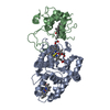
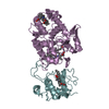
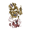
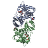
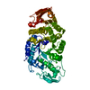
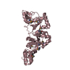
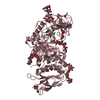
| Title | The SoxAX Complex of Rhodovulum Sulfidophilum | ||||||
 Components Components |
| ||||||
 Keywords Keywords |  ELECTRON TRANSPORT / ELECTRON TRANSFER / BACTERIAL SULFUR CYCLE / THIOSULFATE OXIDATION / CYSTEINE PERSULFIDE HEME LIGAND / CYTOCHROME C ELECTRON TRANSPORT / ELECTRON TRANSFER / BACTERIAL SULFUR CYCLE / THIOSULFATE OXIDATION / CYSTEINE PERSULFIDE HEME LIGAND / CYTOCHROME C | ||||||
| Function / homology |  Function and homology information Function and homology informationL-cysteine S-thiosulfotransferase / oxidoreductase activity, acting on a sulfur group of donors, cytochrome as acceptor / sulfide oxidation / sulfur oxidation / sulfurtransferase activity / cytochrome complex / thiosulfate sulfurtransferase activity / periplasmic space / electron transfer activity / oxidoreductase activity ...L-cysteine S-thiosulfotransferase / oxidoreductase activity, acting on a sulfur group of donors, cytochrome as acceptor / sulfide oxidation / sulfur oxidation / sulfurtransferase activity / cytochrome complex / thiosulfate sulfurtransferase activity / periplasmic space / electron transfer activity / oxidoreductase activity / protein heterodimerization activity / heme binding / metal ion bindingSimilarity search - Function | ||||||
| Biological species |  Rhodovulum sulfidophilum (bacteria) Rhodovulum sulfidophilum (bacteria) | ||||||
| Method | X-RAY DIFFRACTION / SYNCHROTRON / MOLECULAR REPLACEMENT / Resolution: 2.35 Å | ||||||
 Authors Authors | Kihlken, M.A. / Berks, B.C. / Hemmings, A.M. | ||||||
 Citation Citation | Journal: To be Published Title: The crystal structure of recombinant Rhodovulum sulfidophilum SoxAX confirms cysteine persulfide coordination to the catalytic heme Authors: Kihlken, M.A. / Berks, B.C. / Hemmings, A.M. | ||||||
| History |
|
- Structure visualization
| Structure viewer | Molecule: MolmilJmol/JSmol |
|---|
- Downloads & links
-Download
| PDBx/mmCIF format | 2oz1.cif.gz | 341.4 KB | Display | PDBx/mmCIF format |
|---|---|---|---|---|
| PDB format | pdb2oz1.ent.gz | 278.7 KB | Display | PDB format |
| PDBx/mmJSON format | 2oz1.json.gz | Tree view | PDBx/mmJSON format | |
| Others |  Other downloads Other downloads |
-Validation report
| Arichive directory | https://data.pdbj.org/pub/pdb/validation_reports/oz/2oz1ftp://data.pdbj.org/pub/pdb/validation_reports/oz/2oz1 | HTTPS FTP |
|---|
-Related structure data
| Related structure data |  1h33S S: Starting model for refinement |
|---|---|
| Similar structure data |


-Links
 PDBj
PDBj


- Assembly
| Deposited unit | 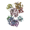
| ||||||||
|---|---|---|---|---|---|---|---|---|---|
| 1 | 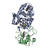
| ||||||||
| 2 | 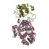
| ||||||||
| 3 | 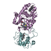
| ||||||||
| 4 | 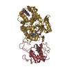
| ||||||||
| Unit cell |
| ||||||||
| Details | THE BIOLOGICAL ASSEMBLY IS A SOXAX HETERODIMER. THE CRYSTALLOGRAPHIC ASYMMETRIC UNIT CONTAINS FOUR INDEPENDENT COPIES OF THE BIOLOGICAL ASSEMBLY. |
-Components
| #1: Protein | Mass: 28974.408 Da / Num. of mol.: 4 Source method: isolated from a genetically manipulated source Source: (gene. exp.) Rhodovulum sulfidophilum (bacteria) / Strain: 1374T / Gene: soxA / Plasmid: pMKS12 / Production host: Rhodobacter capsulatus (bacteria) / Strain (production host): 37B4DELTADORA / References: UniProt: Q939U1#2: Protein | Mass: 14756.748 Da / Num. of mol.: 4 Source method: isolated from a genetically manipulated source Source: (gene. exp.) Rhodovulum sulfidophilum (bacteria) / Strain: 1374T / Gene: soxX / Plasmid: pMKS12 / Production host: Rhodobacter capsulatus (bacteria) / Strain (production host): 37B4DELTADORA / References: UniProt: Q939U4#3: Chemical | ChemComp-HEC / Heme C  Mass: 618.503 Da / Num. of mol.: 12 / Source method: obtained synthetically / Formula: C34H34FeN4O4 Mass: 618.503 Da / Num. of mol.: 12 / Source method: obtained synthetically / Formula: C34H34FeN4O4#4: Water | ChemComp-HOH / | Water Mass: 18.015 Da / Num. of mol.: 939 / Source method: isolated from a natural source / Formula: H2O Mass: 18.015 Da / Num. of mol.: 939 / Source method: isolated from a natural source / Formula: H2O |
|---|
-Experimental details
-Experiment
| Experiment | Method: X-RAY DIFFRACTION / Number of used crystals: 1 |
|---|
- Sample preparation
| Crystal | Density Matthews: 2.59 Å3/Da / Density % sol: 52.43 % |
|---|---|
| Crystal grow | Temperature: 277 K / Method: vapor diffusion, hanging drop / pH: 6.5 Details: 15% (w/v) PEG 4K, 0.2 M magnesium acetate, 100 mM sodium MES buffer pH 6.5. The crystals could be cryoprotected by transferring them to stabilization solution containing 25% (v/v) ethylene ...Details: 15% (w/v) PEG 4K, 0.2 M magnesium acetate, 100 mM sodium MES buffer pH 6.5. The crystals could be cryoprotected by transferring them to stabilization solution containing 25% (v/v) ethylene glycol. , VAPOR DIFFUSION, HANGING DROP, temperature 277K |
-Data collection
| Diffraction | Mean temperature: 100 K | ||||||||||||||||||||||||||||||||||||||||||||||||||||||||||||||||||||||||||||||||||||||||
|---|---|---|---|---|---|---|---|---|---|---|---|---|---|---|---|---|---|---|---|---|---|---|---|---|---|---|---|---|---|---|---|---|---|---|---|---|---|---|---|---|---|---|---|---|---|---|---|---|---|---|---|---|---|---|---|---|---|---|---|---|---|---|---|---|---|---|---|---|---|---|---|---|---|---|---|---|---|---|---|---|---|---|---|---|---|---|---|---|---|
| Diffraction source | Source: SYNCHROTRON / Site: ESRF  / Beamline: ID14-3 / Wavelength: 0.931 Å / Beamline: ID14-3 / Wavelength: 0.931 Å | ||||||||||||||||||||||||||||||||||||||||||||||||||||||||||||||||||||||||||||||||||||||||
| Detector | Type: MAR CCD 165 mm / Detector: CCD / Date: Feb 15, 2005 | ||||||||||||||||||||||||||||||||||||||||||||||||||||||||||||||||||||||||||||||||||||||||
| Radiation | Monochromator: Diamond (111), Ge(220) / Protocol: SINGLE WAVELENGTH / Monochromatic (M) / Laue (L): M / Scattering type: x-ray | ||||||||||||||||||||||||||||||||||||||||||||||||||||||||||||||||||||||||||||||||||||||||
| Radiation wavelength | Wavelength: 0.931 Å / Relative weight: 1 | ||||||||||||||||||||||||||||||||||||||||||||||||||||||||||||||||||||||||||||||||||||||||
| Reflection | Resolution: 2.345→108.577 Å / Num. all: 71803 / Num. obs: 71803 / % possible obs: 96 % / Observed criterion σ(F): 0 / Observed criterion σ(I): 0 / Redundancy: 2.7 % / Biso Wilson estimate: 26.4 Å2 / Rmerge(I) obs: 0.086 / Rsym value: 0.086 / Net I/σ(I): 7.4 | ||||||||||||||||||||||||||||||||||||||||||||||||||||||||||||||||||||||||||||||||||||||||
| Reflection shell |
|
- Processing
| Software |
| ||||||||||||||||||||||||||||||||||||||||||||||||||||||||||||||||||||||||||||||||||||||||||
|---|---|---|---|---|---|---|---|---|---|---|---|---|---|---|---|---|---|---|---|---|---|---|---|---|---|---|---|---|---|---|---|---|---|---|---|---|---|---|---|---|---|---|---|---|---|---|---|---|---|---|---|---|---|---|---|---|---|---|---|---|---|---|---|---|---|---|---|---|---|---|---|---|---|---|---|---|---|---|---|---|---|---|---|---|---|---|---|---|---|---|---|
| Refinement | Method to determine structure: MOLECULAR REPLACEMENT Starting model: PDB ENTRY 1H33 Resolution: 2.35→50 Å / Cor.coef. Fo:Fc: 0.922 / Cor.coef. Fo:Fc free: 0.868 / SU B: 7.203 / SU ML: 0.175 / Isotropic thermal model: INDIVIDUAL ISOTROPIC / Cross valid method: THROUGHOUT / σ(F): 0 / σ(I): 0 / ESU R: 0.455 / ESU R Free: 0.262 / Stereochemistry target values: MAXIMUM LIKELIHOOD / Details: HYDROGENS HAVE BEEN ADDED IN THE RIDING POSITIONS
| ||||||||||||||||||||||||||||||||||||||||||||||||||||||||||||||||||||||||||||||||||||||||||
| Solvent computation | Ion probe radii: 0.8 Å / Shrinkage radii: 0.8 Å / VDW probe radii: 1.2 Å / Solvent model: MASK | ||||||||||||||||||||||||||||||||||||||||||||||||||||||||||||||||||||||||||||||||||||||||||
| Displacement parameters | Biso mean: 14.774 Å2
| ||||||||||||||||||||||||||||||||||||||||||||||||||||||||||||||||||||||||||||||||||||||||||
| Refinement step | Cycle: LAST / Resolution: 2.35→50 Å
| ||||||||||||||||||||||||||||||||||||||||||||||||||||||||||||||||||||||||||||||||||||||||||
| Refine LS restraints |
| ||||||||||||||||||||||||||||||||||||||||||||||||||||||||||||||||||||||||||||||||||||||||||
| LS refinement shell | Resolution: 2.345→2.406 Å / Total num. of bins used: 20
|
