 Movie
Movie Controller


+ Open data

- Basic information
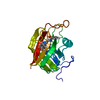
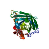
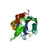
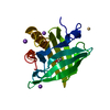
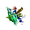
| Entry | Database: PDB / ID: 1xyh | ||||||
|---|---|---|---|---|---|---|---|
| Title | Crystal Structure of Recombinant Human Cyclophilin J | ||||||
 Components Components | cyclophilin-like protein PPIL3b | ||||||
 Keywords Keywords |  ISOMERASE / cyclophilin J / beta-barrel / helix / disulfide bridge ISOMERASE / cyclophilin J / beta-barrel / helix / disulfide bridge | ||||||
| Function / homology |  Function and homology information Function and homology informationcatalytic step 2 spliceosome / mRNA Splicing - Major Pathway / peptidylprolyl isomerase / peptidyl-prolyl cis-trans isomerase activity / mRNA splicing, via spliceosome / protein folding / nucleoplasmSimilarity search - Function | ||||||
| Biological species |  Homo sapiens (human) Homo sapiens (human) | ||||||
| Method | X-RAY DIFFRACTION / MOLECULAR REPLACEMENT / Resolution: 2.6 Å | ||||||
 Authors Authors | Huang, L.-L. / Zhao, X.-M. / Huang, C.-Q. / Yu, L. / Xia, Z.-X. | ||||||
 Citation Citation | Journal: Acta Crystallogr.,Sect.D / Year: 2005 Title: Structure of recombinant human cyclophilin J, a novel member of the cyclophilin family. Authors: Huang, L.L. / Zhao, X.M. / Huang, C.Q. / Yu, L. / Xia, Z.X. | ||||||
| History |
| ||||||
| Remark 650 | HELIX DETERMINATION METHOD: AUTHOR DETERMINED | ||||||
| Remark 700 | SHEET DETERMINATION METHOD: AUTHOR DETERMINED |
- Structure visualization
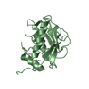
| Structure viewer | Molecule: MolmilJmol/JSmol |
|---|
- Downloads & links
-Download
| PDBx/mmCIF format | 1xyh.cif.gz | 44.1 KB | Display | PDBx/mmCIF format |
|---|---|---|---|---|
| PDB format | pdb1xyh.ent.gz | 30.3 KB | Display | PDB format |
| PDBx/mmJSON format | 1xyh.json.gz | Tree view | PDBx/mmJSON format | |
| Others |  Other downloads Other downloads |
-Validation report
| Arichive directory | https://data.pdbj.org/pub/pdb/validation_reports/xy/1xyhftp://data.pdbj.org/pub/pdb/validation_reports/xy/1xyh | HTTPS FTP |
|---|
-Related structure data
| Related structure data |  2cplS S: Starting model for refinement |
|---|---|
| Similar structure data |

-Links
 PDBj
PDBj


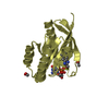
- Assembly
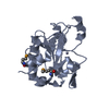
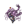
| Deposited unit | 
| ||||||||
|---|---|---|---|---|---|---|---|---|---|
| 1 |
| ||||||||
| Unit cell |
|
-Components
| #1: Protein | Mass: 18177.547 Da / Num. of mol.: 1 Source method: isolated from a genetically manipulated source Source: (gene. exp.) Homo sapiens (human) / Gene: PPIL3b / Organ: fetal brain / Plasmid: pTXB1 / Species (production host): Escherichia coli / Production host:  Escherichia coli BL21 (bacteria) / Strain (production host): BL21 / References: UniProt: Q9H2H8, peptidylprolyl isomerase Escherichia coli BL21 (bacteria) / Strain (production host): BL21 / References: UniProt: Q9H2H8, peptidylprolyl isomerase |
|---|---|
| #2: Water | ChemComp-HOH / Water Mass: 18.015 Da / Num. of mol.: 26 / Source method: isolated from a natural source / Formula: H2O Mass: 18.015 Da / Num. of mol.: 26 / Source method: isolated from a natural source / Formula: H2O |
-Experimental details
-Experiment
| Experiment | Method: X-RAY DIFFRACTION / Number of used crystals: 1 |
|---|
- Sample preparation
| Crystal | Density Matthews: 2.35 Å3/Da / Density % sol: 47.7 % |
|---|---|
| Crystal grow | Temperature: 293 K / Method: vapor diffusion, hanging drop / pH: 7.6 Details: Tris-HCl, PEG8000, DMSO, pH 7.6, VAPOR DIFFUSION, HANGING DROP, temperature 293.0K |
-Data collection
| Diffraction | Mean temperature: 293 K |
|---|---|
| Diffraction source | Source: SEALED TUBE / Type: OTHER / Wavelength: 1.5418 Å |
| Detector | Type: BRUKER / Detector: CCD / Date: Jun 26, 2004 |
| Radiation | Monochromator: graphite / Protocol: SINGLE WAVELENGTH / Monochromatic (M) / Laue (L): M / Scattering type: x-ray |
| Radiation wavelength | Wavelength: 1.5418 Å / Relative weight: 1 |
| Reflection | Resolution: 2.6→27.51 Å / Num. all: 5798 / Num. obs: 5560 / % possible obs: 95.9 % / Observed criterion σ(I): 0 / Biso Wilson estimate: 30.8 Å2 / Rmerge(I) obs: 0.084 |
| Reflection shell | Resolution: 2.6→2.69 Å / Rmerge(I) obs: 0.372 / Num. unique all: 499 / % possible all: 92.4 |
- Processing
| Software |
| ||||||||||||||||||||||||||||||||||||
|---|---|---|---|---|---|---|---|---|---|---|---|---|---|---|---|---|---|---|---|---|---|---|---|---|---|---|---|---|---|---|---|---|---|---|---|---|---|
| Refinement | Method to determine structure: MOLECULAR REPLACEMENT Starting model: PDB ENTRY 2CPL Resolution: 2.6→27.51 Å / Rfactor Rfree error: 0.01 / Data cutoff high absF: 227854.65 / Data cutoff low absF: 0 / Isotropic thermal model: RESTRAINED / Cross valid method: THROUGHOUT / σ(F): 0 Stereochemistry target values: maximum likelyhood target using amplitude
| ||||||||||||||||||||||||||||||||||||
| Solvent computation | Solvent model: FLAT MODEL / Bsol: 26.4574 Å2 / ksol: 0.301138 e/Å3 | ||||||||||||||||||||||||||||||||||||
| Displacement parameters | Biso mean: 28.4 Å2
| ||||||||||||||||||||||||||||||||||||
| Refine analyze |
| ||||||||||||||||||||||||||||||||||||
| Refinement step | Cycle: LAST / Resolution: 2.6→27.51 Å
| ||||||||||||||||||||||||||||||||||||
| Refine LS restraints |
| ||||||||||||||||||||||||||||||||||||
| LS refinement shell | Resolution: 2.6→2.69 Å / Rfactor Rfree error: 0.051 / Total num. of bins used: 10
| ||||||||||||||||||||||||||||||||||||
| Xplor file |
|
