 Movie
Movie Controller

[English] 日本語
 Yorodumi
- PDB-3hyq: Crystal Structure of Isopentenyl-Diphosphate delta-Isomerase from... -
+ Open data

- Basic information
| Entry | Database: PDB / ID: 3hyq | ||||||
|---|---|---|---|---|---|---|---|
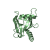
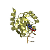
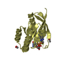
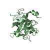
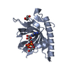
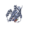
| Title | Crystal Structure of Isopentenyl-Diphosphate delta-Isomerase from Salmonella entericase | ||||||
 Components Components | Isopentenyl-diphosphate Delta-isomerase Isopentenyl-diphosphate delta isomerase Isopentenyl-diphosphate delta isomerase | ||||||
 Keywords Keywords | ISOMERASE / alpha-beta structure / Isoprene biosynthesis / Magnesium / Manganese / Metal-binding / Structural Genomics / Center for Structural Genomics of Infectious Diseases / CSGID | ||||||
| Function / homology |  Function and homology informationisopentenyl-diphosphate Delta-isomerase / isopentenyl-diphosphate delta-isomerase activity / dimethylallyl diphosphate biosynthetic process / isoprenoid biosynthetic process / hydrolase activity / metal ion binding / cytoplasm Function and homology informationisopentenyl-diphosphate Delta-isomerase / isopentenyl-diphosphate delta-isomerase activity / dimethylallyl diphosphate biosynthetic process / isoprenoid biosynthetic process / hydrolase activity / metal ion binding / cytoplasmSimilarity search - Function | ||||||
| Biological species |  Salmonella enterica subsp. enterica serovar Typhimurium (bacteria) Salmonella enterica subsp. enterica serovar Typhimurium (bacteria) | ||||||
| Method | X-RAY DIFFRACTION / SYNCHROTRON / SAD / Resolution: 1.525 Å | ||||||
 Authors Authors | Kim, Y. / Zhou, M. / Peterson, S. / Anderson, W.F. / Joachimiak, A. / Center for Structural Genomics of Infectious Diseases (CSGID) | ||||||
 Citation Citation | Journal: To be Published Title: Crystal Structure of Isopentenyl-Diphosphate delta-Isomerase from Salmonella entericase Authors: Kim, Y. / Zhou, M. / Peterson, S. / Anderson, W.F. / Joachimiak, A. | ||||||
| History |
|
- Structure visualization
| Structure viewer | Molecule: MolmilJmol/JSmol |
|---|
- Downloads & links
-Download
| PDBx/mmCIF format | 3hyq.cif.gz | 49.9 KB | Display | PDBx/mmCIF format |
|---|---|---|---|---|
| PDB format | pdb3hyq.ent.gz | 38.8 KB | Display | PDB format |
| PDBx/mmJSON format | 3hyq.json.gz | Tree view | PDBx/mmJSON format | |
| Others |  Other downloads Other downloads |
-Validation report
| Arichive directory | https://data.pdbj.org/pub/pdb/validation_reports/hy/3hyqftp://data.pdbj.org/pub/pdb/validation_reports/hy/3hyq | HTTPS FTP |
|---|
-Related structure data
| Similar structure data | |
|---|---|
| Other databases |


-Links
 PDBj
PDBj

- Assembly
| Deposited unit | 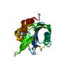
| |||||||||
|---|---|---|---|---|---|---|---|---|---|---|
| 1 |
| |||||||||
| 2 | 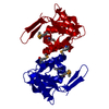
| |||||||||
| Unit cell |
| |||||||||
| Components on special symmetry positions |
| |||||||||
| Details | monomer |
-Components
| #1: Protein | Isopentenyl-diphosphate delta isomerase / IPP isomerase / Isopentenyl pyrophosphate isomerase / IPP:DMAPP isomerase Mass: 21262.264 Da / Num. of mol.: 1 Source method: isolated from a genetically manipulated source Source: (gene. exp.) Salmonella enterica subsp. enterica serovar Typhimurium (bacteria)Strain: LT2 / Gene: idi / Plasmid: pMCSG7 / Production host: Escherichia coli (E. coli) / Strain (production host): BL21 magicReferences: UniProt: Q8ZM82, isopentenyl-diphosphate Delta-isomerase |
|---|---|
| #2: Water | ChemComp-HOH / Water Mass: 18.015 Da / Num. of mol.: 227 / Source method: isolated from a natural source / Formula: H2O Mass: 18.015 Da / Num. of mol.: 227 / Source method: isolated from a natural source / Formula: H2O |
-Experimental details
-Experiment
| Experiment | Method: X-RAY DIFFRACTION / Number of used crystals: 1 |
|---|
- Sample preparation
| Crystal | Density Matthews: 2.12 Å3/Da / Density % sol: 41.96 % |
|---|---|
| Crystal grow | Temperature: 289 K / Method: vapor diffusion, sitting drop Details: 0.4 M ammonium dihydrogen phosphate, chymotripsyn (1:100), VAPOR DIFFUSION, SITTING DROP, temperature 289K |
-Data collection
| Diffraction | Mean temperature: 100 K |
|---|---|
| Diffraction source | Source: SYNCHROTRON / Site: APS  / Beamline: 19-BM / Wavelength: 0.9792 Å / Beamline: 19-BM / Wavelength: 0.9792 Å |
| Detector | Type: ADSC QUANTUM 210r / Detector: CCD / Date: May 31, 2009 / Details: mirrors |
| Radiation | Monochromator: double crystal monochromator / Protocol: SAD / Monochromatic (M) / Laue (L): M / Scattering type: x-ray |
| Radiation wavelength | Wavelength: 0.9792 Å / Relative weight: 1 |
| Reflection | Resolution: 1.53→50 Å / Num. all: 26450 / Num. obs: 26450 / % possible obs: 98.1 % / Observed criterion σ(F): 0 / Observed criterion σ(I): 0 / Redundancy: 3.9 % / Biso Wilson estimate: 21.74 Å2 / Rsym value: 0.062 / Net I/σ(I): 19 |
| Reflection shell | Resolution: 1.53→1.56 Å / Redundancy: 2 % / Mean I/σ(I) obs: 2.1 / Num. unique all: 1015 / Rsym value: 0.308 / % possible all: 78 |
- Processing
| Software |
| ||||||||||||||||||||||||||||||||||||||||||||||||||||||||||||||||||||||
|---|---|---|---|---|---|---|---|---|---|---|---|---|---|---|---|---|---|---|---|---|---|---|---|---|---|---|---|---|---|---|---|---|---|---|---|---|---|---|---|---|---|---|---|---|---|---|---|---|---|---|---|---|---|---|---|---|---|---|---|---|---|---|---|---|---|---|---|---|---|---|---|
| Refinement | Method to determine structure: SAD / Resolution: 1.525→25.005 Å / SU ML: 0.09 / Cross valid method: THROUGHOUT / σ(F): 0 / Stereochemistry target values: MLHL
| ||||||||||||||||||||||||||||||||||||||||||||||||||||||||||||||||||||||
| Solvent computation | Shrinkage radii: 0.9 Å / VDW probe radii: 1.11 Å / Solvent model: FLAT BULK SOLVENT MODEL / Bsol: 59.683 Å2 / ksol: 0.362 e/Å3 | ||||||||||||||||||||||||||||||||||||||||||||||||||||||||||||||||||||||
| Displacement parameters |
| ||||||||||||||||||||||||||||||||||||||||||||||||||||||||||||||||||||||
| Refinement step | Cycle: LAST / Resolution: 1.525→25.005 Å
| ||||||||||||||||||||||||||||||||||||||||||||||||||||||||||||||||||||||
| Refine LS restraints |
| ||||||||||||||||||||||||||||||||||||||||||||||||||||||||||||||||||||||
| LS refinement shell | Refine-ID: X-RAY DIFFRACTION
|
