 Movie
Movie Controller

[English] 日本語
 Yorodumi
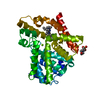
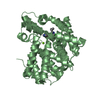
- PDB-1xp0: Catalytic Domain Of Human Phosphodiesterase 5A In Complex With Va... -
+ Open data

- Basic information
| Entry | Database: PDB / ID: 1xp0 | ||||||
|---|---|---|---|---|---|---|---|
| Title | Catalytic Domain Of Human Phosphodiesterase 5A In Complex With Vardenafil | ||||||
 Components Components | cGMP-specific 3',5'-cyclic phosphodiesterase | ||||||
 Keywords Keywords |  HYDROLASE / Phosphodiesterase / PDE / PDE5A / Vardenafil / Levitra HYDROLASE / Phosphodiesterase / PDE / PDE5A / Vardenafil / Levitra | ||||||
| Function / homology |  Function and homology information Function and homology informationpositive regulation of oocyte development / 3',5'-cyclic-GMP phosphodiesterase / regulation of nitric oxide mediated signal transduction / negative regulation of cardiac muscle contraction / oocyte development / RHOBTB1 GTPase cycle / relaxation of cardiac muscle / positive regulation of cardiac muscle hypertrophy / cGMP catabolic process / cGMP effects ...positive regulation of oocyte development / 3',5'-cyclic-GMP phosphodiesterase / regulation of nitric oxide mediated signal transduction / negative regulation of cardiac muscle contraction / oocyte development / RHOBTB1 GTPase cycle / relaxation of cardiac muscle / positive regulation of cardiac muscle hypertrophy / cGMP catabolic process / cGMP effects / cGMP binding / 3',5'-cyclic-nucleotide phosphodiesterase activity / 3',5'-cyclic-GMP phosphodiesterase activity / Smooth Muscle Contraction / T cell proliferation / negative regulation of T cell proliferation / signal transduction / metal ion binding / cytosolSimilarity search - Function | ||||||
| Biological species |  Homo sapiens (human) Homo sapiens (human) | ||||||
| Method | X-RAY DIFFRACTION / SYNCHROTRON / MOLECULAR REPLACEMENT / Resolution: 1.79 Å | ||||||
 Authors Authors | Card, G.L. / England, B.P. / Suzuki, Y. / Fong, D. / Powell, B. / Lee, B. / Luu, C. / Tabrizizad, M. / Gillette, S. / Ibrahim, P.N. ...Card, G.L. / England, B.P. / Suzuki, Y. / Fong, D. / Powell, B. / Lee, B. / Luu, C. / Tabrizizad, M. / Gillette, S. / Ibrahim, P.N. / Artis, D.R. / Bollag, G. / Milburn, M.V. / Kim, S.-H. / Schlessinger, J. / Zhang, K.Y.J. | ||||||
 Citation Citation | Journal: STRUCTURE / Year: 2004 Title: Structural Basis for the Activity of Drugs that Inhibit Phosphodiesterases. Authors: Card, G.L. / England, B.P. / Suzuki, Y. / Fong, D. / Powell, B. / Lee, B. / Luu, C. / Tabrizizad, M. / Gillette, S. / Ibrahim, P.N. / Artis, D.R. / Bollag, G. / Milburn, M.V. / Kim, S.-H. / ...Authors: Card, G.L. / England, B.P. / Suzuki, Y. / Fong, D. / Powell, B. / Lee, B. / Luu, C. / Tabrizizad, M. / Gillette, S. / Ibrahim, P.N. / Artis, D.R. / Bollag, G. / Milburn, M.V. / Kim, S.-H. / Schlessinger, J. / Zhang, K.Y.J. | ||||||
| History |
| ||||||
| Remark 600 | HETEROGEN HOH 1001-1007 ARE ASSOCIATED WITH CHAIN A. |
- Structure visualization
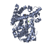
| Structure viewer | Molecule: MolmilJmol/JSmol |
|---|
- Downloads & links
-Download
| PDBx/mmCIF format | 1xp0.cif.gz | 85.3 KB | Display | PDBx/mmCIF format |
|---|---|---|---|---|
| PDB format | pdb1xp0.ent.gz | 63.3 KB | Display | PDB format |
| PDBx/mmJSON format | 1xp0.json.gz | Tree view | PDBx/mmJSON format | |
| Others |  Other downloads Other downloads |
-Validation report
| Arichive directory | https://data.pdbj.org/pub/pdb/validation_reports/xp/1xp0ftp://data.pdbj.org/pub/pdb/validation_reports/xp/1xp0 | HTTPS FTP |
|---|
-Related structure data
| Related structure data | 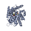 1xlxC 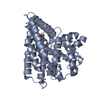 1xlzC 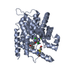 1xm4C 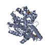 1xm6C 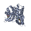 1xmuC 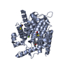 1xmyC 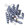 1xn0C 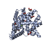 1xomC 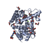 1xonC 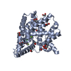 1xoqC 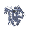 1xorC 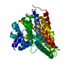 1xosC 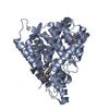 1xotC 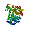 1xozC C: citing same article ( |
|---|---|
| Similar structure data |


-Links
 PDBj
PDBj


- Assembly
| Deposited unit | 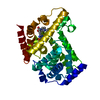
| ||||||||
|---|---|---|---|---|---|---|---|---|---|
| 1 |
| ||||||||
| Unit cell |
| ||||||||
| Details | The biological assembly is one monomer. |
-Components
| #1: Protein | Mass: 41827.934 Da / Num. of mol.: 1 / Fragment: CATALYTIC DOMAIN OF HUMAN PHOSPHODIESTERASE 5A Source method: isolated from a genetically manipulated source Source: (gene. exp.) Homo sapiens (human) / Gene: PDE5A, PDE5 / Plasmid: pET15b / Production host:  Escherichia coli (E. coli) / Strain (production host): BL21(DE3)Codon Plus(RIL) Escherichia coli (E. coli) / Strain (production host): BL21(DE3)Codon Plus(RIL)References: UniProt: O76074, 3',5'-cyclic-nucleotide phosphodiesterase |
|---|---|
| #2: Chemical | ChemComp-ZN /   Mass: 65.409 Da / Num. of mol.: 1 / Source method: obtained synthetically / Formula: Zn Mass: 65.409 Da / Num. of mol.: 1 / Source method: obtained synthetically / Formula: Zn |
| #3: Chemical | ChemComp-MG /   Mass: 24.305 Da / Num. of mol.: 1 / Source method: obtained synthetically / Formula: Mg Mass: 24.305 Da / Num. of mol.: 1 / Source method: obtained synthetically / Formula: Mg |
| #4: Chemical | ChemComp-VDN / Vardenafil  Mass: 488.603 Da / Num. of mol.: 1 / Source method: obtained synthetically / Formula: C23H32N6O4S / Comment: medication, inhibitor*YM Mass: 488.603 Da / Num. of mol.: 1 / Source method: obtained synthetically / Formula: C23H32N6O4S / Comment: medication, inhibitor*YM |
| #5: Water | ChemComp-HOH / Water Mass: 18.015 Da / Num. of mol.: 156 / Source method: isolated from a natural source / Formula: H2O Mass: 18.015 Da / Num. of mol.: 156 / Source method: isolated from a natural source / Formula: H2O |
-Experimental details
-Experiment
| Experiment | Method: X-RAY DIFFRACTION / Number of used crystals: 1 |
|---|
- Sample preparation
| Crystal | Density Matthews: 3.08 Å3/Da / Density % sol: 60.05 % |
|---|---|
| Crystal grow | Temperature: 277 K / Method: vapor diffusion, sitting drop / pH: 7 Details: Sodium formate, pH 7.0, VAPOR DIFFUSION, SITTING DROP, temperature 277K |
-Data collection
| Diffraction | Mean temperature: 93 K |
|---|---|
| Diffraction source | Source: SYNCHROTRON / Site: ALS  / Beamline: 8.3.1 / Wavelength: 1.1 Å / Beamline: 8.3.1 / Wavelength: 1.1 Å |
| Detector | Type: ADSC QUANTUM 210 / Detector: CCD / Date: Sep 4, 2003 |
| Radiation | Protocol: SINGLE WAVELENGTH / Monochromatic (M) / Laue (L): M / Scattering type: x-ray |
| Radiation wavelength | Wavelength: 1.1 Å / Relative weight: 1 |
| Reflection | Resolution: 1.79→76.7 Å / Num. all: 46654 / Num. obs: 46654 / % possible obs: 96.9 % / Observed criterion σ(F): 0 / Observed criterion σ(I): 0 / Redundancy: 6 % / Rmerge(I) obs: 0.058 / Net I/σ(I): 7.8 |
| Reflection shell | Resolution: 1.79→1.836 Å / Redundancy: 5.5 % / Rmerge(I) obs: 0.599 / Mean I/σ(I) obs: 1.3 / Num. unique all: 3490 / % possible all: 96.9 |
- Processing
| Software |
| ||||||||||||||||||||||||||||||||||||||||||||||||||||||||||||||||||||||||||||||||||||||||||||||||||||||||||||||||||||||||||||||||||||||||||||||||||||||||||||||||
|---|---|---|---|---|---|---|---|---|---|---|---|---|---|---|---|---|---|---|---|---|---|---|---|---|---|---|---|---|---|---|---|---|---|---|---|---|---|---|---|---|---|---|---|---|---|---|---|---|---|---|---|---|---|---|---|---|---|---|---|---|---|---|---|---|---|---|---|---|---|---|---|---|---|---|---|---|---|---|---|---|---|---|---|---|---|---|---|---|---|---|---|---|---|---|---|---|---|---|---|---|---|---|---|---|---|---|---|---|---|---|---|---|---|---|---|---|---|---|---|---|---|---|---|---|---|---|---|---|---|---|---|---|---|---|---|---|---|---|---|---|---|---|---|---|---|---|---|---|---|---|---|---|---|---|---|---|---|---|---|---|---|
| Refinement | Method to determine structure: MOLECULAR REPLACEMENT / Resolution: 1.79→76.7 Å / Cor.coef. Fo:Fc: 0.966 / Cor.coef. Fo:Fc free: 0.958 / SU B: 2.921 / SU ML: 0.083 / TLS residual ADP flag: LIKELY RESIDUAL / Isotropic thermal model: Isotropic / Cross valid method: THROUGHOUT / ESU R: 0.101 / ESU R Free: 0.094 / Stereochemistry target values: MAXIMUM LIKELIHOOD / Details: HYDROGENS HAVE BEEN ADDED IN THE RIDING POSITIONS
| ||||||||||||||||||||||||||||||||||||||||||||||||||||||||||||||||||||||||||||||||||||||||||||||||||||||||||||||||||||||||||||||||||||||||||||||||||||||||||||||||
| Solvent computation | Ion probe radii: 0.8 Å / Shrinkage radii: 0.8 Å / VDW probe radii: 1.4 Å / Solvent model: BABINET MODEL WITH MASK | ||||||||||||||||||||||||||||||||||||||||||||||||||||||||||||||||||||||||||||||||||||||||||||||||||||||||||||||||||||||||||||||||||||||||||||||||||||||||||||||||
| Displacement parameters | Biso mean: 20.466 Å2
| ||||||||||||||||||||||||||||||||||||||||||||||||||||||||||||||||||||||||||||||||||||||||||||||||||||||||||||||||||||||||||||||||||||||||||||||||||||||||||||||||
| Refine analyze |
| ||||||||||||||||||||||||||||||||||||||||||||||||||||||||||||||||||||||||||||||||||||||||||||||||||||||||||||||||||||||||||||||||||||||||||||||||||||||||||||||||
| Refinement step | Cycle: LAST / Resolution: 1.79→76.7 Å
| ||||||||||||||||||||||||||||||||||||||||||||||||||||||||||||||||||||||||||||||||||||||||||||||||||||||||||||||||||||||||||||||||||||||||||||||||||||||||||||||||
| Refine LS restraints |
| ||||||||||||||||||||||||||||||||||||||||||||||||||||||||||||||||||||||||||||||||||||||||||||||||||||||||||||||||||||||||||||||||||||||||||||||||||||||||||||||||
| LS refinement shell | Resolution: 1.79→1.836 Å / Total num. of bins used: 20 /
| ||||||||||||||||||||||||||||||||||||||||||||||||||||||||||||||||||||||||||||||||||||||||||||||||||||||||||||||||||||||||||||||||||||||||||||||||||||||||||||||||
| Refinement TLS params. | Method: refined / Origin x: -24.341 Å / Origin y: 39.855 Å / Origin z: 70.624 Å
|
