 Movie
Movie Controller

[English] 日本語
 Yorodumi
- PDB-1xhd: X-ray crystal structure of putative acetyltransferase, product of... -
+ Open data

- Basic information
| Entry | Database: PDB / ID: 1xhd | ||||||
|---|---|---|---|---|---|---|---|
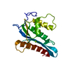
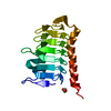
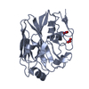
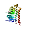
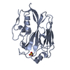
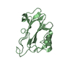
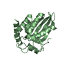
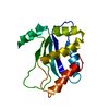
| Title | X-ray crystal structure of putative acetyltransferase, product of BC4754 gene [Bacillus cereus] | ||||||
 Components Components | putative acetyltransferase/acyltransferase | ||||||
 Keywords Keywords |  TRANSFERASE / structural genomics / protein structure initiative / Medwest Center for Structural Genomics / MCSG / Macyltransferase / PSI / Midwest Center for Structural Genomics TRANSFERASE / structural genomics / protein structure initiative / Medwest Center for Structural Genomics / MCSG / Macyltransferase / PSI / Midwest Center for Structural Genomics | ||||||
| Function / homology | Hexapeptide repeat proteins / UDP N-Acetylglucosamine Acyltransferase; domain 1 / Hexapeptide repeat / Bacterial transferase hexapeptide (six repeats) / acyltransferase activity / Trimeric LpxA-like superfamily / 3 Solenoid / Mainly Beta / Putative acetyltransferase/acyltransferase Function and homology information Function and homology information | ||||||
| Biological species |  Bacillus cereus (bacteria) Bacillus cereus (bacteria) | ||||||
| Method | X-RAY DIFFRACTION / SYNCHROTRON / SAD / Resolution: 1.9 Å | ||||||
 Authors Authors | Osipiuk, J. / Zhou, M. / Moy, S. / Collart, F. / Joachimiak, A. / Midwest Center for Structural Genomics (MCSG) | ||||||
 Citation Citation | Journal: To be Published Title: X-ray crystal structure of putative acetyltransferase, product of BC4754 gene from Bacillus cereus. Authors: Osipiuk, J. / Zhou, M. / Moy, S. / Collart, F. / Joachimiak, A. / Midwest Center for Structural Genomics (MCSG) | ||||||
| History |
| ||||||
| Remark 300 | BIOMOLECULE: 1 THIS ENTRY CONTAINS THE CRYSTALLOGRAPHIC ASYMMETRIC UNIT WHICH CONSISTS OF 1 CHAIN(S) ...BIOMOLECULE: 1 THIS ENTRY CONTAINS THE CRYSTALLOGRAPHIC ASYMMETRIC UNIT WHICH CONSISTS OF 1 CHAIN(S). THE BIOLOGICAL MOLECULE FOR THE PROTEIN IS UNKNOWN. |
- Structure visualization
| Structure viewer | Molecule: MolmilJmol/JSmol |
|---|
- Downloads & links
-Download
| PDBx/mmCIF format | 1xhd.cif.gz | 48 KB | Display | PDBx/mmCIF format |
|---|---|---|---|---|
| PDB format | pdb1xhd.ent.gz | 37.1 KB | Display | PDB format |
| PDBx/mmJSON format | 1xhd.json.gz | Tree view | PDBx/mmJSON format | |
| Others |  Other downloads Other downloads |
-Validation report
| Arichive directory | https://data.pdbj.org/pub/pdb/validation_reports/xh/1xhdftp://data.pdbj.org/pub/pdb/validation_reports/xh/1xhd | HTTPS FTP |
|---|
-Related structure data
| Similar structure data | |
|---|---|
| Other databases |


-Links
 PDBj
PDBj


- Assembly
| Deposited unit | 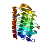
| |||||||||
|---|---|---|---|---|---|---|---|---|---|---|
| 1 |
| |||||||||
| 2 | 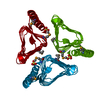
| |||||||||
| Unit cell |
| |||||||||
| Components on special symmetry positions |
| |||||||||
| Details | the biological assembly unknown |
-Components
| #1: Protein | Mass: 19277.531 Da / Num. of mol.: 1 Source method: isolated from a genetically manipulated source Source: (gene. exp.) Bacillus cereus (bacteria) / Strain: ATCC 14579 / Gene: BC4754 / Plasmid: pMCSG7 / Production host: Escherichia coli (E. coli) / Strain (production host): BL21[DE3]pMAGIC / References: UniProt: Q816R4 |
|---|---|
| #2: Chemical | ChemComp-SO4 / Sulfate  Mass: 96.063 Da / Num. of mol.: 1 / Source method: obtained synthetically / Formula: SO4 Mass: 96.063 Da / Num. of mol.: 1 / Source method: obtained synthetically / Formula: SO4 |
| #3: Water | ChemComp-HOH / Water Mass: 18.015 Da / Num. of mol.: 130 / Source method: isolated from a natural source / Formula: H2O Mass: 18.015 Da / Num. of mol.: 130 / Source method: isolated from a natural source / Formula: H2O |
-Experimental details
-Experiment
| Experiment | Method: X-RAY DIFFRACTION / Number of used crystals: 1 |
|---|
- Sample preparation
| Crystal | Density Matthews: 6.3 Å3/Da / Density % sol: 80.2 % |
|---|---|
| Crystal grow | Temperature: 289 K / Method: vapor diffusion, sitting drop / pH: 4.2 Details: SODIUM/POTASSIUM PHOSPHATE, PHOSPHATE CITRATE, pH 4.2, VAPOR DIFFUSION, SITTING DROP, temperature 289K |
-Data collection
| Diffraction | Mean temperature: 100 K |
|---|---|
| Diffraction source | Source: SYNCHROTRON / Site: APS  / Beamline: 19-ID / Wavelength: 0.979295 Å / Beamline: 19-ID / Wavelength: 0.979295 Å |
| Detector | Type: SBC-3 / Detector: CCD / Date: Jun 16, 2004 |
| Radiation | Monochromator: double crystal monochromator / Protocol: SINGLE WAVELENGTH / Monochromatic (M) / Laue (L): M / Scattering type: x-ray |
| Radiation wavelength | Wavelength: 0.979295 Å / Relative weight: 1 |
| Reflection | Resolution: 1.9→40 Å / Num. all: 37577 / Num. obs: 37525 / % possible obs: 99.9 % / Observed criterion σ(F): 0 / Observed criterion σ(I): 0 / Redundancy: 41.5 % / Rmerge(I) obs: 0.101 / Net I/σ(I): 48.4 |
| Reflection shell | Resolution: 1.9→1.94 Å / Redundancy: 15.7 % / Rmerge(I) obs: 0.794 / Mean I/σ(I) obs: 2.49 / Num. unique all: 2401 / % possible all: 98.1 |
- Processing
| Software |
| ||||||||||||||||||||||||||||||||||||||||||||||||||||||||||||||||||||||||||||||||||||||||||||||||||||||||||||||||||||||||||||||||||||||||||||||||||||||||||||||||
|---|---|---|---|---|---|---|---|---|---|---|---|---|---|---|---|---|---|---|---|---|---|---|---|---|---|---|---|---|---|---|---|---|---|---|---|---|---|---|---|---|---|---|---|---|---|---|---|---|---|---|---|---|---|---|---|---|---|---|---|---|---|---|---|---|---|---|---|---|---|---|---|---|---|---|---|---|---|---|---|---|---|---|---|---|---|---|---|---|---|---|---|---|---|---|---|---|---|---|---|---|---|---|---|---|---|---|---|---|---|---|---|---|---|---|---|---|---|---|---|---|---|---|---|---|---|---|---|---|---|---|---|---|---|---|---|---|---|---|---|---|---|---|---|---|---|---|---|---|---|---|---|---|---|---|---|---|---|---|---|---|---|
| Refinement | Method to determine structure: SAD / Resolution: 1.9→40 Å / Cor.coef. Fo:Fc: 0.97 / SU B: 1.098 / SU ML: 0.033 / TLS residual ADP flag: LIKELY RESIDUAL / σ(F): 0 / ESU R: 0.063 / Stereochemistry target values: MAXIMUM LIKELIHOOD / Details: HYDROGENS HAVE BEEN ADDED IN THE RIDING POSITIONS
| ||||||||||||||||||||||||||||||||||||||||||||||||||||||||||||||||||||||||||||||||||||||||||||||||||||||||||||||||||||||||||||||||||||||||||||||||||||||||||||||||
| Solvent computation | Ion probe radii: 0.8 Å / Shrinkage radii: 0.8 Å / VDW probe radii: 1.4 Å / Solvent model: BABINET MODEL WITH MASK | ||||||||||||||||||||||||||||||||||||||||||||||||||||||||||||||||||||||||||||||||||||||||||||||||||||||||||||||||||||||||||||||||||||||||||||||||||||||||||||||||
| Displacement parameters | Biso mean: 19.555 Å2 | ||||||||||||||||||||||||||||||||||||||||||||||||||||||||||||||||||||||||||||||||||||||||||||||||||||||||||||||||||||||||||||||||||||||||||||||||||||||||||||||||
| Refinement step | Cycle: LAST / Resolution: 1.9→40 Å
| ||||||||||||||||||||||||||||||||||||||||||||||||||||||||||||||||||||||||||||||||||||||||||||||||||||||||||||||||||||||||||||||||||||||||||||||||||||||||||||||||
| Refine LS restraints |
| ||||||||||||||||||||||||||||||||||||||||||||||||||||||||||||||||||||||||||||||||||||||||||||||||||||||||||||||||||||||||||||||||||||||||||||||||||||||||||||||||
| LS refinement shell | Resolution: 1.9→1.949 Å / Total num. of bins used: 20
| ||||||||||||||||||||||||||||||||||||||||||||||||||||||||||||||||||||||||||||||||||||||||||||||||||||||||||||||||||||||||||||||||||||||||||||||||||||||||||||||||
| Refinement TLS params. | Method: refined / Origin x: -8.5637 Å / Origin y: 26.0727 Å / Origin z: 98.1143 Å
|
