 Movie
Movie Controller

[English] 日本語
 Yorodumi
- PDB-1xg6: The crystal structure of the P1 mutant (Leu to Arg)of a Winged be... -
+ Open data

- Basic information
| Entry | Database: PDB / ID: 1xg6 | ||||||
|---|---|---|---|---|---|---|---|
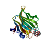
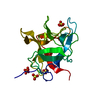
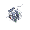
| Title | The crystal structure of the P1 mutant (Leu to Arg)of a Winged bean chymotrypsin inhibitor(Kunitz)solved at 2.15A resolution | ||||||
 Components Components | Chymotrypsin inhibitor 3 | ||||||
 Keywords Keywords | Hydrolase Inhibitor /  trypsin inhibitor trypsin inhibitor | ||||||
| Function / homology |  Function and homology information Function and homology information | ||||||
| Biological species |   Psophocarpus tetragonolobus (winged bean) Psophocarpus tetragonolobus (winged bean) | ||||||
| Method | X-RAY DIFFRACTION / MOLECULAR REPLACEMENT / Resolution: 2.15 Å | ||||||
 Authors Authors | Sen, U. / Dattagupta, J.K. / Dasgupta, J. / Khamrui, S. | ||||||
 Citation Citation | Journal: Biochim.Biophys.Acta / Year: 2005 Title: Single mutation at P1 of a chymotrypsin inhibitor changes it to a trypsin inhibitor: X-ray structural (2.15 A) and biochemical basis Authors: Khamrui, S. / Dasgupta, J. / Dattagupta, J.K. / Sen, U. #1: Journal: Proteins / Year: 1999Title: Refined crystal structure (2.3 A) of a double-headed winged bean alpha-chymotrypsin inhibitor and location of its second reactive site Authors: Dattagupta, J.K. / Podder, A. / Chakrabarti, C. / Sen, U. / Mukhopadhyay, D. / Dutta, S.K. / Singh, M. #2: Journal: Protein Eng. / Year: 2001Title: The role of Asn14 in the stability and conformation of the reactive-site loop of winged bean chymotrypsin inhibitor: crystal structures of two point mutants Asn14-->Lys and Asn14-->Asp Authors: Ravichandran, S. / Dasgupta, J. / Chakrabarti, C. / Ghosh, S. / Singh, M. / Dattagupta, J.K. | ||||||
| History |
|
- Structure visualization
| Structure viewer | Molecule: MolmilJmol/JSmol |
|---|
- Downloads & links
-Download
| PDBx/mmCIF format | 1xg6.cif.gz | 53.3 KB | Display | PDBx/mmCIF format |
|---|---|---|---|---|
| PDB format | pdb1xg6.ent.gz | 37.4 KB | Display | PDB format |
| PDBx/mmJSON format | 1xg6.json.gz | Tree view | PDBx/mmJSON format | |
| Others |  Other downloads Other downloads |
-Validation report
| Arichive directory | https://data.pdbj.org/pub/pdb/validation_reports/xg/1xg6ftp://data.pdbj.org/pub/pdb/validation_reports/xg/1xg6 | HTTPS FTP |
|---|
-Related structure data
| Related structure data |  1eylS S: Starting model for refinement |
|---|---|
| Similar structure data |


-Links
 PDBj
PDBj- Assembly
| Deposited unit | 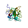
| ||||||||
|---|---|---|---|---|---|---|---|---|---|
| 1 |
| ||||||||
| Unit cell |
|
-Components
| #1: Protein | Mass: 20724.357 Da / Num. of mol.: 1 / Mutation: L68R Source method: isolated from a genetically manipulated source Source: (gene. exp.) Psophocarpus tetragonolobus (winged bean)Plasmid: pET28a+ / Production host:  Escherichia coli (E. coli) / References: UniProt: P10822 Escherichia coli (E. coli) / References: UniProt: P10822 |
|---|---|
| #2: Water | ChemComp-HOH / Water Mass: 18.015 Da / Num. of mol.: 190 / Source method: isolated from a natural source / Formula: H2O Mass: 18.015 Da / Num. of mol.: 190 / Source method: isolated from a natural source / Formula: H2O |
-Experimental details
-Experiment
| Experiment | Method: X-RAY DIFFRACTION / Number of used crystals: 1 |
|---|
- Sample preparation
| Crystal | Density Matthews: 2.48 Å3/Da / Density % sol: 52.19 % |
|---|
-Data collection
| Diffraction | Mean temperature: 100 K |
|---|---|
| Diffraction source | Source: ROTATING ANODE / Type: RIGAKU RU200 / Wavelength: 1.5418 |
| Detector | Type: MARRESEARCH / Detector: IMAGE PLATE / Date: Jun 6, 2004 / Details: Osmic MaxFlux |
| Radiation | Monochromator: Copper / Protocol: SINGLE WAVELENGTH / Monochromatic (M) / Laue (L): M / Scattering type: x-ray |
| Radiation wavelength | Wavelength: 1.5418 Å / Relative weight: 1 |
| Reflection | Resolution: 2.15→15 Å / Num. obs: 11202 / Biso Wilson estimate: 29.3 Å2 |
- Processing
| Software |
| ||||||||||||||||||||||||||||||||||||
|---|---|---|---|---|---|---|---|---|---|---|---|---|---|---|---|---|---|---|---|---|---|---|---|---|---|---|---|---|---|---|---|---|---|---|---|---|---|
| Refinement | Method to determine structure: MOLECULAR REPLACEMENT Starting model: 1EYL Resolution: 2.15→14.93 Å / Rfactor Rfree error: 0.01 / Data cutoff high absF: 1249123.49 / Data cutoff low absF: 0 / Isotropic thermal model: RESTRAINED / Cross valid method: THROUGHOUT / σ(F): 0 / Stereochemistry target values: Engh & Huber
| ||||||||||||||||||||||||||||||||||||
| Solvent computation | Solvent model: FLAT MODEL / Bsol: 53.5872 Å2 / ksol: 0.318978 e/Å3 | ||||||||||||||||||||||||||||||||||||
| Displacement parameters | Biso mean: 45.9 Å2
| ||||||||||||||||||||||||||||||||||||
| Refine analyze |
| ||||||||||||||||||||||||||||||||||||
| Refinement step | Cycle: LAST / Resolution: 2.15→14.93 Å
| ||||||||||||||||||||||||||||||||||||
| Refine LS restraints |
| ||||||||||||||||||||||||||||||||||||
| LS refinement shell | Resolution: 2.15→2.28 Å / Rfactor Rfree error: 0.041 / Total num. of bins used: 6
| ||||||||||||||||||||||||||||||||||||
| Xplor file |
|
