 Movie
Movie Controller

[English] 日本語
 Yorodumi
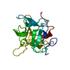
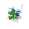
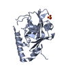
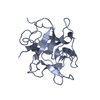
- PDB-1fmz: CRYSTAL STRUCTURE OF A MUTANT WINGED BEAN CHYMOTRYPSIN INHIBITOR ... -
+ Open data

- Basic information
| Entry | Database: PDB / ID: 1fmz | ||||||
|---|---|---|---|---|---|---|---|
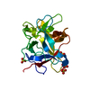
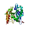
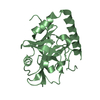
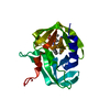
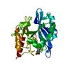
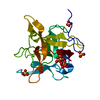
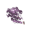
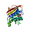
| Title | CRYSTAL STRUCTURE OF A MUTANT WINGED BEAN CHYMOTRYPSIN INHIBITOR PROTEIN, N14K. | ||||||
 Components Components | CHYMOTRYPSIN INHIBITOR 3 | ||||||
 Keywords Keywords | HYDROLASE INHIBITOR / Beta Trefoil | ||||||
| Function / homology |  Function and homology information Function and homology information | ||||||
| Biological species |    Psophocarpus tetragonolobus (winged bean) Psophocarpus tetragonolobus (winged bean) | ||||||
| Method | X-RAY DIFFRACTION / SYNCHROTRON / Resolution: 2.05 Å | ||||||
 Authors Authors | Dattagupta, J.K. / Chakrabarti, C. / Ravichandran, S. / Dasgupta, J. / Ghosh, S. | ||||||
 Citation Citation | Journal: PROTEIN ENG. / Year: 2001 Title: The role of Asn14 in the stability and conformation of the reactive-site loop of winged bean chymotrypsin inhibitor: crystal structures of two point mutants Asn14-->Lys and Asn14-->Asp. Authors: Ravichandran, S. / Dasgupta, J. / Chakrabarti, C. / Ghosh, S. / Singh, M. / Dattagupta, J.K. #1: Journal: Acta Crystallogr.,Sect.D / Year: 1999Title: Crystallography of a Kunitz-type Serine Protease Inhibitor: The 90K Structure of Winged bean Chymotrypsin Inhibitor (WCI) at 2.13A resolution. Authors: Ravichandran, S. / Sen, U. / Chakrabarti, C. / Dattagupta, J.K. #2: Journal: Proteins / Year: 1999Title: Refined crystal structure (2.3A) of a double-headed Winged bean alpha-Chymotrypsin Inhibitor and location of its second reactive site Authors: Dattagupta, J.K. / Podder, A. / Chakrabarti, C. / Sen, U. / Mukhopadhyay, D. / Dutta, S.K. / Singh, M. #3: Journal: Protein Expr.Purif. / Year: 1997Title: cDNA cloning, expression and rapid purification of Kunitz-type Winged bean Chymotrypsin Inhibitor Authors: Ghosh, S. / Singh, M. | ||||||
| History |
|
- Structure visualization
| Structure viewer | Molecule: MolmilJmol/JSmol |
|---|
- Downloads & links
-Download
| PDBx/mmCIF format | 1fmz.cif.gz | 50.3 KB | Display | PDBx/mmCIF format |
|---|---|---|---|---|
| PDB format | pdb1fmz.ent.gz | 39.6 KB | Display | PDB format |
| PDBx/mmJSON format | 1fmz.json.gz | Tree view | PDBx/mmJSON format | |
| Others |  Other downloads Other downloads |
-Validation report
| Arichive directory | https://data.pdbj.org/pub/pdb/validation_reports/fm/1fmzftp://data.pdbj.org/pub/pdb/validation_reports/fm/1fmz | HTTPS FTP |
|---|
-Related structure data


-Links
 PDBj
PDBj- Assembly
| Deposited unit | 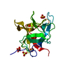
| ||||||||||||
|---|---|---|---|---|---|---|---|---|---|---|---|---|---|
| 1 |
| ||||||||||||
| 2 | 
| ||||||||||||
| 3 | 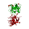
| ||||||||||||
| Unit cell |
| ||||||||||||
| Components on special symmetry positions |
| ||||||||||||
| Details | The biological assembly is a monomer |
-Components
| #1: Protein | Mass: 20689.408 Da / Num. of mol.: 1 / Mutation: N14K Source method: isolated from a genetically manipulated source Source: (gene. exp.) Psophocarpus tetragonolobus (winged bean)Organ: SEED / Plasmid: PTRC99A / Production host:  Escherichia coli (E. coli) / References: UniProt: P10822 Escherichia coli (E. coli) / References: UniProt: P10822 | ||
|---|---|---|---|
| #2: Chemical | ChemComp-SO4 / Sulfate  Mass: 96.063 Da / Num. of mol.: 5 / Source method: obtained synthetically / Formula: SO4 Mass: 96.063 Da / Num. of mol.: 5 / Source method: obtained synthetically / Formula: SO4#3: Water | ChemComp-HOH / | Water Mass: 18.015 Da / Num. of mol.: 179 / Source method: isolated from a natural source / Formula: H2O Mass: 18.015 Da / Num. of mol.: 179 / Source method: isolated from a natural source / Formula: H2O |
-Experimental details
-Experiment
| Experiment | Method: X-RAY DIFFRACTION / Number of used crystals: 1 |
|---|
- Sample preparation
| Crystal | Density Matthews: 2.69 Å3/Da / Density % sol: 54.3 % | ||||||||||||||||||||||||||||||||||||||||||||||||
|---|---|---|---|---|---|---|---|---|---|---|---|---|---|---|---|---|---|---|---|---|---|---|---|---|---|---|---|---|---|---|---|---|---|---|---|---|---|---|---|---|---|---|---|---|---|---|---|---|---|
| Crystal grow | Temperature: 277 K / Method: vapor diffusion, hanging drop / pH: 5.4 Details: Ammonium sulfate, Sodium acetate, pH 5.4, VAPOR DIFFUSION, HANGING DROP, temperature 277.0K | ||||||||||||||||||||||||||||||||||||||||||||||||
| Crystal grow | *PLUS Temperature: 4 ℃Details: Dattagupta, J.K., (1999) Proteins: Struct., Funct., Genet., 35, 321. | ||||||||||||||||||||||||||||||||||||||||||||||||
| Components of the solutions | *PLUS
|
-Data collection
| Diffraction | Mean temperature: 100 K |
|---|---|
| Diffraction source | Source: SYNCHROTRON / Site: ESRF  / Beamline: ID14-4 / Wavelength: 0.934 / Beamline: ID14-4 / Wavelength: 0.934 |
| Detector | Type: MARRESEARCH / Detector: CCD / Date: Jul 22, 1998 |
| Radiation | Protocol: SINGLE WAVELENGTH / Monochromatic (M) / Laue (L): M / Scattering type: x-ray |
| Radiation wavelength | Wavelength: 0.934 Å / Relative weight: 1 |
| Reflection | Resolution: 2.05→15 Å / Num. all: 147330 / Num. obs: 13051 / % possible obs: 88.1 % / Observed criterion σ(F): 3 / Observed criterion σ(I): 3 / Redundancy: 4.3 % / Biso Wilson estimate: 28.1 Å2 / Rmerge(I) obs: 0.082 / Net I/σ(I): 5.2 |
| Reflection shell | Resolution: 2.05→2.16 Å / Redundancy: 5.4 % / Rmerge(I) obs: 0.282 / % possible all: 88.1 |
| Reflection | *PLUS Num. measured all: 147330 |
| Reflection shell | *PLUS % possible obs: 91.8 % |
- Processing
| Software |
| ||||||||||||||||||||||||||||||
|---|---|---|---|---|---|---|---|---|---|---|---|---|---|---|---|---|---|---|---|---|---|---|---|---|---|---|---|---|---|---|---|
| Refinement | Resolution: 2.05→15 Å / σ(F): 0 / σ(I): 0 / Stereochemistry target values: Engh & Huber / Details: Used Maximum likelihood target function
| ||||||||||||||||||||||||||||||
| Refinement step | Cycle: LAST / Resolution: 2.05→15 Å
| ||||||||||||||||||||||||||||||
| Software | *PLUS Name: REFMAC / Classification: refinement | ||||||||||||||||||||||||||||||
| Refinement | *PLUS σ(F): 0 / Rfactor all: 0.21 / Rfactor obs: 0.193 | ||||||||||||||||||||||||||||||
| Solvent computation | *PLUS | ||||||||||||||||||||||||||||||
| Displacement parameters | *PLUS | ||||||||||||||||||||||||||||||
| Refine LS restraints | *PLUS
|
