 Movie
Movie Controller

[English] 日本語
 Yorodumi
- PDB-2wbc: REFINED CRYSTAL STRUCTURE (2.3 ANGSTROM) OF A WINGED BEAN CHYMOTR... -
+ Open data

- Basic information
| Entry | Database: PDB / ID: 2wbc | ||||||
|---|---|---|---|---|---|---|---|
| Title | REFINED CRYSTAL STRUCTURE (2.3 ANGSTROM) OF A WINGED BEAN CHYMOTRYPSIN INHIBITOR AND LOCATION OF ITS SECOND REACTIVE SITE | ||||||
 Components Components | CHYMOTRYPSIN INHIBITOR | ||||||
 Keywords Keywords |  SERINE PROTEASE INHIBITOR SERINE PROTEASE INHIBITOR | ||||||
| Function / homology |  Function and homology information Function and homology information | ||||||
| Biological species |   Psophocarpus tetragonolobus (winged bean) Psophocarpus tetragonolobus (winged bean) | ||||||
| Method | X-RAY DIFFRACTION / Resolution: 2.3 Å | ||||||
 Authors Authors | Dattagupta, J.K. / Podder, A. / Chakrabarti, C. / Sen, U. / Mukhopadhyay, D. / Dutta, S.K. / Singh, M. | ||||||
 Citation Citation | Journal: Proteins / Year: 1999 Title: Refined crystal structure (2.3 A) of a double-headed winged bean alpha-chymotrypsin inhibitor and location of its second reactive site. Authors: Dattagupta, J.K. / Podder, A. / Chakrabarti, C. / Sen, U. / Mukhopadhyay, D. / Dutta, S.K. / Singh, M. #1: Journal: J.Mol.Biol. / Year: 1990Title: Crystallization and Preliminary X-Ray Studies of Psophocarpin B1, a Chymotrypsin Inhibitor from Winged Bean Seeds Authors: Dattagupta, J.K. / Chakrabarti, C. / Podder, A. / Dutta, S.K. / Singh, M. | ||||||
| History |
|
- Structure visualization
| Structure viewer | Molecule: MolmilJmol/JSmol |
|---|
- Downloads & links
-Download
| PDBx/mmCIF format | 2wbc.cif.gz | 56.9 KB | Display | PDBx/mmCIF format |
|---|---|---|---|---|
| PDB format | pdb2wbc.ent.gz | 46.2 KB | Display | PDB format |
| PDBx/mmJSON format | 2wbc.json.gz | Tree view | PDBx/mmJSON format | |
| Others |  Other downloads Other downloads |
-Validation report
| Arichive directory | https://data.pdbj.org/pub/pdb/validation_reports/wb/2wbcftp://data.pdbj.org/pub/pdb/validation_reports/wb/2wbc | HTTPS FTP |
|---|
-Related structure data
| Similar structure data |
|---|


-Links
 PDBj
PDBj- Assembly
| Deposited unit | 
| ||||||||
|---|---|---|---|---|---|---|---|---|---|
| 1 |
| ||||||||
| Unit cell |
|
-Components
| #1: Protein | Mass: 20266.850 Da / Num. of mol.: 1 / Source method: isolated from a natural source / Source: (natural) Psophocarpus tetragonolobus (winged bean) / Tissue: SEED / References: UniProt: P10822 |
|---|---|
| #2: Water | ChemComp-HOH / Water Mass: 18.015 Da / Num. of mol.: 109 / Source method: isolated from a natural source / Formula: H2O Mass: 18.015 Da / Num. of mol.: 109 / Source method: isolated from a natural source / Formula: H2O |
-Experimental details
-Experiment
| Experiment | Method: X-RAY DIFFRACTION / Number of used crystals: 1 |
|---|
- Sample preparation
| Crystal | Density Matthews: 3.85 Å3/Da / Density % sol: 68 % | ||||||||||||||||||||||||||||||||||||||||||||||||
|---|---|---|---|---|---|---|---|---|---|---|---|---|---|---|---|---|---|---|---|---|---|---|---|---|---|---|---|---|---|---|---|---|---|---|---|---|---|---|---|---|---|---|---|---|---|---|---|---|---|
| Crystal grow | Temperature: 277 K / Method: vapor diffusion, hanging drop / pH: 5.4 Details: PROTEIN WAS CRYSTALLIZED BY HANGING DROP VAPOUR DIFFUSION FROM 25% AMM. SULPHATE IN TRIS-HCL, 10MM NA ACETATE,400 MM NACL AT PH 5.4 AGAINST 25% AMM. SULPHATE,60MM NA ACETATE AT 4 DEGREE ...Details: PROTEIN WAS CRYSTALLIZED BY HANGING DROP VAPOUR DIFFUSION FROM 25% AMM. SULPHATE IN TRIS-HCL, 10MM NA ACETATE,400 MM NACL AT PH 5.4 AGAINST 25% AMM. SULPHATE,60MM NA ACETATE AT 4 DEGREE CENTIGRADE., vapor diffusion - hanging drop, temperature 277K | ||||||||||||||||||||||||||||||||||||||||||||||||
| Crystal | *PLUS | ||||||||||||||||||||||||||||||||||||||||||||||||
| Crystal grow | *PLUS Temperature: 4 ℃ / Method: vapor diffusion, hanging drop | ||||||||||||||||||||||||||||||||||||||||||||||||
| Components of the solutions | *PLUS
|
-Data collection
| Diffraction | Mean temperature: 293 K |
|---|---|
| Diffraction source | Source: ROTATING ANODE / Type: RIGAKU FR-D / Wavelength: 1.5418 |
| Detector | Type: RIGAKU / Detector: IMAGE PLATE / Date: May 1, 1996 / Details: MIRRORS |
| Radiation | Monochromator: NI FILTER / Monochromatic (M) / Laue (L): M / Scattering type: x-ray |
| Radiation wavelength | Wavelength: 1.5418 Å / Relative weight: 1 |
| Reflection | Resolution: 2.3→12 Å / Num. obs: 9897 / % possible obs: 80.1 % / Observed criterion σ(I): 1 / Redundancy: 3.1 % / Biso Wilson estimate: 34.2 Å2 / Rmerge(I) obs: 0.095 |
| Reflection | *PLUS Num. measured all: 10551 |
- Processing
| Software |
| ||||||||||||||||||||||||||||||||||||||||||||||||||||||||||||
|---|---|---|---|---|---|---|---|---|---|---|---|---|---|---|---|---|---|---|---|---|---|---|---|---|---|---|---|---|---|---|---|---|---|---|---|---|---|---|---|---|---|---|---|---|---|---|---|---|---|---|---|---|---|---|---|---|---|---|---|---|---|
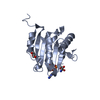
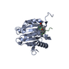
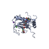
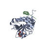
| Refinement | Resolution: 2.3→8 Å / Isotropic thermal model: RESTRAINED / Cross valid method: THROUGHOUT / σ(F): 2 Details: THE ELECTRON DENSITY IS CONTINUOUS FOR ALL THE MAIN CHAIN ATOMS. HOWEVER THE FITTING OF THE MODEL TO THE ELECTRON DENSITY MAP IN THE RESIDUE RANGES 137 - 142, AND 176 - 183 IS SOMEWHAT POOR. ...Details: THE ELECTRON DENSITY IS CONTINUOUS FOR ALL THE MAIN CHAIN ATOMS. HOWEVER THE FITTING OF THE MODEL TO THE ELECTRON DENSITY MAP IN THE RESIDUE RANGES 137 - 142, AND 176 - 183 IS SOMEWHAT POOR. WCI HAS BEEN FOUND TO BE SPHERICAL AND CONSISTS OF 12 ANTIPARALLEL BETA-STRANDS WITH CONNECTING LOOPS ARRANGED IN A CHARACTERISTIC B-TREFOIL TYPE FOLDING. A PSEUDO THREE-FOLD AXIS EXISTS PARALLEL TO THE BARREL AXIS OF THE STRUCTURE. EACH OF THE THREE SUBDOMAINS COMPRISES FOUR B-STRANDS WITH CONNECTING LOOPS. FROM THIS HIGHER RESOLUTION MODEL THE LOCATION OF THE SECOND REACTIVE SITE OF THE INHIBITOR PROTEIN COULD BE IDENTIFIED MAINLY ON THE BASIS OF STRUCTURAL STUDIES.
| ||||||||||||||||||||||||||||||||||||||||||||||||||||||||||||
| Displacement parameters | Biso mean: 30.8 Å2 | ||||||||||||||||||||||||||||||||||||||||||||||||||||||||||||
| Refine analyze | Luzzati coordinate error obs: 0.28 Å / Luzzati d res low obs: 8 Å | ||||||||||||||||||||||||||||||||||||||||||||||||||||||||||||
| Refinement step | Cycle: LAST / Resolution: 2.3→8 Å
| ||||||||||||||||||||||||||||||||||||||||||||||||||||||||||||
| Refine LS restraints |
| ||||||||||||||||||||||||||||||||||||||||||||||||||||||||||||
| Xplor file |
| ||||||||||||||||||||||||||||||||||||||||||||||||||||||||||||
| Software | *PLUS Name: X-PLOR / Version: 3.1 / Classification: refinement | ||||||||||||||||||||||||||||||||||||||||||||||||||||||||||||
| Refine LS restraints | *PLUS
|
