 Movie
Movie Controller

[English] 日本語
 Yorodumi
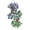
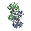
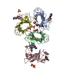
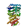
- PDB-1xdu: Crystal structure of Aclacinomycin-10-hydroxylase (RdmB) in compl... -
+ Open data

- Basic information
| Entry | Database: PDB / ID: 1xdu | ||||||
|---|---|---|---|---|---|---|---|
| Title | Crystal structure of Aclacinomycin-10-hydroxylase (RdmB) in complex with Sinefungin (SFG) | ||||||
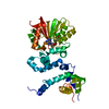
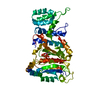
 Components Components | Protein RdmB | ||||||
 Keywords Keywords |  TRANSFERASE / Anthracycline / hydroxylase / S-adenosyl-L-methionine analogue / sinefungin / streptomyces / polyketide antibiotics / divergent evolution TRANSFERASE / Anthracycline / hydroxylase / S-adenosyl-L-methionine analogue / sinefungin / streptomyces / polyketide antibiotics / divergent evolution | ||||||
| Function / homology |  Function and homology informationLyases; Carbon-carbon lyases; Carboxy-lyases / O-methyltransferase activity / carboxy-lyase activity / antibiotic biosynthetic process Function and homology informationLyases; Carbon-carbon lyases; Carboxy-lyases / O-methyltransferase activity / carboxy-lyase activity / antibiotic biosynthetic processSimilarity search - Function | ||||||
| Biological species |  Streptomyces purpurascens (bacteria) Streptomyces purpurascens (bacteria) | ||||||
| Method | X-RAY DIFFRACTION / SYNCHROTRON / MOLECULAR REPLACEMENT / Resolution: 2.7 Å | ||||||
 Authors Authors | Jansson, A. / Koskiniemi, H. / Erola, A. / Wang, J. / Mantsala, P. / Schneider, G. / Niemi, J. | ||||||
 Citation Citation | Journal: J.Biol.Chem. / Year: 2005 Title: Aclacinomycin 10-Hydroxylase Is a Novel Substrate-assisted Hydroxylase Requiring S-Adenosyl-L-methionine as Cofactor Authors: Jansson, A. / Koskiniemi, H. / Erola, A. / Wang, J. / Schneider, G. / Niemi, J. | ||||||
| History |
|
- Structure visualization
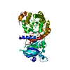
| Structure viewer | Molecule: MolmilJmol/JSmol |
|---|
- Downloads & links
-Download
| PDBx/mmCIF format | 1xdu.cif.gz | 79 KB | Display | PDBx/mmCIF format |
|---|---|---|---|---|
| PDB format | pdb1xdu.ent.gz | 58.8 KB | Display | PDB format |
| PDBx/mmJSON format | 1xdu.json.gz | Tree view | PDBx/mmJSON format | |
| Others |  Other downloads Other downloads |
-Validation report
| Arichive directory | https://data.pdbj.org/pub/pdb/validation_reports/xd/1xduftp://data.pdbj.org/pub/pdb/validation_reports/xd/1xdu | HTTPS FTP |
|---|
-Related structure data


-Links
 PDBj
PDBj
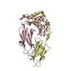
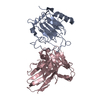
- Assembly
| Deposited unit | 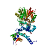
| ||||||||||
|---|---|---|---|---|---|---|---|---|---|---|---|
| 1 |
| ||||||||||
| 2 | 
| ||||||||||
| Unit cell |
|
-Components
| #1: Protein | Mass: 40166.207 Da / Num. of mol.: 1 Source method: isolated from a genetically manipulated source Source: (gene. exp.) Streptomyces purpurascens (bacteria) / Gene: rdmb / Plasmid: pRDM16 from pGEX4T-3 / Production host: Escherichia coli (E. coli) / References: UniProt: Q54527 |
|---|---|
| #2: Chemical | ChemComp-ACT / Acetate  Mass: 59.044 Da / Num. of mol.: 1 / Source method: obtained synthetically / Formula: C2H3O2 Mass: 59.044 Da / Num. of mol.: 1 / Source method: obtained synthetically / Formula: C2H3O2 |
| #3: Chemical | ChemComp-SFG /   Mass: 381.387 Da / Num. of mol.: 1 / Source method: obtained synthetically / Formula: C15H23N7O5 Mass: 381.387 Da / Num. of mol.: 1 / Source method: obtained synthetically / Formula: C15H23N7O5 |
| #4: Water | ChemComp-HOH / Water Mass: 18.015 Da / Num. of mol.: 26 / Source method: isolated from a natural source / Formula: H2O Mass: 18.015 Da / Num. of mol.: 26 / Source method: isolated from a natural source / Formula: H2O |
-Experimental details
-Experiment
| Experiment | Method: X-RAY DIFFRACTION / Number of used crystals: 1 |
|---|
- Sample preparation
| Crystal | Density Matthews: 2.17 Å3/Da / Density % sol: 43.7 % |
|---|---|
| Crystal grow | Temperature: 294 K / Method: vapor diffusion, hanging drop / pH: 4.6 Details: ammonium acetate, PEG 4000, sodium acetate, pH 4.6, VAPOR DIFFUSION, HANGING DROP, temperature 294K |
-Data collection
| Diffraction | Mean temperature: 100 K |
|---|---|
| Diffraction source | Source: SYNCHROTRON / Site: ESRF  / Beamline: ID29 / Wavelength: 1.05 Å / Beamline: ID29 / Wavelength: 1.05 Å |
| Detector | Type: ADSC QUANTUM 4 / Detector: CCD / Date: Nov 27, 2003 |
| Radiation | Protocol: SINGLE WAVELENGTH / Monochromatic (M) / Laue (L): M / Scattering type: x-ray |
| Radiation wavelength | Wavelength: 1.05 Å / Relative weight: 1 |
| Reflection | Resolution: 2.7→58.7 Å / Num. all: 8940 / Num. obs: 8940 / % possible obs: 98.6 % / Observed criterion σ(F): 0 / Observed criterion σ(I): 0 / Redundancy: 9.82 % / Biso Wilson estimate: 40 Å2 / Rsym value: 0.104 / Net I/σ(I): 12.2 |
| Reflection shell | Resolution: 2.7→2.83 Å / Mean I/σ(I) obs: 4.6 / Rsym value: 0.252 / % possible all: 99.5 |
- Processing
| Software |
| ||||||||||||||||||||||||||||||||||||||||||||||||||||||||||||||||||||||||||||||||||||||||||||||||||||
|---|---|---|---|---|---|---|---|---|---|---|---|---|---|---|---|---|---|---|---|---|---|---|---|---|---|---|---|---|---|---|---|---|---|---|---|---|---|---|---|---|---|---|---|---|---|---|---|---|---|---|---|---|---|---|---|---|---|---|---|---|---|---|---|---|---|---|---|---|---|---|---|---|---|---|---|---|---|---|---|---|---|---|---|---|---|---|---|---|---|---|---|---|---|---|---|---|---|---|---|---|---|
| Refinement | Method to determine structure: MOLECULAR REPLACEMENT Starting model: RdmB+SAM complex Resolution: 2.7→58.72 Å / Cor.coef. Fo:Fc: 0.912 / Cor.coef. Fo:Fc free: 0.854 / SU B: 16.587 / SU ML: 0.343 / Isotropic thermal model: Isotropic B-factors for each atom / Cross valid method: THROUGHOUT / ESU R Free: 0.451 / Stereochemistry target values: MAXIMUM LIKELIHOOD / Details: HYDROGENS HAVE BEEN ADDED IN THE RIDING POSITIONS
| ||||||||||||||||||||||||||||||||||||||||||||||||||||||||||||||||||||||||||||||||||||||||||||||||||||
| Solvent computation | Ion probe radii: 0.8 Å / Shrinkage radii: 0.8 Å / VDW probe radii: 1.4 Å / Solvent model: BABINET MODEL WITH MASK | ||||||||||||||||||||||||||||||||||||||||||||||||||||||||||||||||||||||||||||||||||||||||||||||||||||
| Displacement parameters | Biso mean: 32.176 Å2
| ||||||||||||||||||||||||||||||||||||||||||||||||||||||||||||||||||||||||||||||||||||||||||||||||||||
| Refinement step | Cycle: LAST / Resolution: 2.7→58.72 Å
| ||||||||||||||||||||||||||||||||||||||||||||||||||||||||||||||||||||||||||||||||||||||||||||||||||||
| Refine LS restraints |
| ||||||||||||||||||||||||||||||||||||||||||||||||||||||||||||||||||||||||||||||||||||||||||||||||||||
| LS refinement shell | Resolution: 2.7→2.77 Å / Total num. of bins used: 20 /
|
