Resolution: 2.5→2.54 Å / Redundancy: 6 % / Mean I/σ(I) obs: 2.5 / Rsym value: 0.67 / % possible all: 100
-
Processing
Software
Name
Version
Classification
REFMAC
5.1.24
refinement
HKL-2000
datareduction
HKL-2000
datascaling
SHARP
phasing
SHELXS
phasing
Refinement
Method to determine structure: MAD / Resolution: 2.5→84.51 Å / Cor.coef. Fo:Fc: 0.938 / Cor.coef. Fo:Fc free: 0.938 / SU B: 12.616 / SU ML: 0.266 / TLS residual ADP flag: LIKELY RESIDUAL / Cross valid method: THROUGHOUT / σ(F): 1 / ESU R: 0.433 / ESU R Free: 0.294 Stereochemistry target values: MAXIMUM LIKELIHOOD WITH PHASES Details: HYDROGENS HAVE BEEN ADDED IN THE RIDING POSITIONS. BUSTER-TNT (AUTHOR: G.BRICOGNE) WAS ALSO USED IN THE REFINEMENT.
Rfactor
Num. reflection
% reflection
Selection details
Rfree
0.29147
523
4.8 %
RANDOM
Rwork
0.2634
-
-
-
all
0.2647
10841
-
-
obs
0.26471
10318
99.45 %
-
Solvent computation
Ion probe radii: 0.8 Å / Shrinkage radii: 0.8 Å / VDW probe radii: 1.4 Å / Solvent model: BABINET MODEL WITH MASK
Displacement parameters
Biso mean: 39.135 Å2
Refinement step
Cycle: LAST / Resolution: 2.5→84.51 Å
Protein
Nucleic acid
Ligand
Solvent
Total
Num. atoms
1609
0
45
11
1665
Refine LS restraints
Refine-ID
Type
Dev ideal
Number
X-RAY DIFFRACTION
r_bond_refined_d
0.023
1643
X-RAY DIFFRACTION
r_bond_other_d
0.001
1500
X-RAY DIFFRACTION
r_angle_refined_deg
1.991
2229
X-RAY DIFFRACTION
r_angle_other_deg
0.871
3472
X-RAY DIFFRACTION
r_dihedral_angle_1_deg
6.87
194
X-RAY DIFFRACTION
r_dihedral_angle_2_deg
X-RAY DIFFRACTION
r_dihedral_angle_3_deg
X-RAY DIFFRACTION
r_dihedral_angle_4_deg
X-RAY DIFFRACTION
r_chiral_restr
0.121
230
X-RAY DIFFRACTION
r_gen_planes_refined
0.021
1807
X-RAY DIFFRACTION
r_gen_planes_other
0.007
345
X-RAY DIFFRACTION
r_nbd_refined
0.331
522
X-RAY DIFFRACTION
r_nbd_other
0.292
1971
X-RAY DIFFRACTION
r_nbtor_refined
X-RAY DIFFRACTION
r_nbtor_other
0.105
1036
X-RAY DIFFRACTION
r_xyhbond_nbd_refined
0.283
62
X-RAY DIFFRACTION
r_xyhbond_nbd_other
X-RAY DIFFRACTION
r_metal_ion_refined
0.256
2
X-RAY DIFFRACTION
r_metal_ion_other
X-RAY DIFFRACTION
r_symmetry_vdw_refined
0.167
12
X-RAY DIFFRACTION
r_symmetry_vdw_other
0.294
34
X-RAY DIFFRACTION
r_symmetry_hbond_refined
0.056
4
X-RAY DIFFRACTION
r_symmetry_hbond_other
X-RAY DIFFRACTION
r_mcbond_it
1.573
970
X-RAY DIFFRACTION
r_mcbond_other
X-RAY DIFFRACTION
r_mcangle_it
2.811
1558
X-RAY DIFFRACTION
r_scbond_it
4.885
673
X-RAY DIFFRACTION
r_scangle_it
7.833
671
X-RAY DIFFRACTION
r_rigid_bond_restr
X-RAY DIFFRACTION
r_sphericity_free
X-RAY DIFFRACTION
r_sphericity_bonded
Refinement TLS params.
Method: refined / Refine-ID: X-RAY DIFFRACTION
ID
L11 (°2)
L12 (°2)
L13 (°2)
L22 (°2)
L23 (°2)
L33 (°2)
S11 (Å °)
S12 (Å °)
S13 (Å °)
S21 (Å °)
S22 (Å °)
S23 (Å °)
S31 (Å °)
S32 (Å °)
S33 (Å °)
T11 (Å2)
T12 (Å2)
T13 (Å2)
T22 (Å2)
T23 (Å2)
T33 (Å2)
Origin x (Å)
Origin y (Å)
Origin z (Å)
1
10.7211
0.0056
-0.0012
4.5839
0.7431
4.0555
-0.0562
-0.8772
0.4591
1.9036
0.3065
-0.9605
-0.2319
0.2567
-0.2504
0.9596
0.145
-0.2283
0.7686
0.0735
0.5046
87.189
56.554
25.726
2
9.645
0.8632
-1.196
3.1809
1.012
2.2831
0.0179
-0.9599
-0.1814
0.9529
0.1513
0.3971
0.0549
0.0673
-0.1692
0.2206
0.121
0.0325
0.3768
0.2039
0.1164
69.444
54.35
14.84
Refinement TLS group
ID
Refine-ID
Refine TLS-ID
Auth asym-ID
Auth seq-ID
1
X-RAY DIFFRACTION
1
A
1 - 95
2
X-RAY DIFFRACTION
2
A
96 - 195
+
About Yorodumi
-
News
-
Feb 9, 2022. New format data for meta-information of EMDB entries
New format data for meta-information of EMDB entries
Version 3 of the EMDB header file is now the official format.
The previous official version 1.9 will be removed from the archive.
In the structure databanks used in Yorodumi, some data are registered as the other names, "COVID-19 virus" and "2019-nCoV". Here are the details of the virus and the list of structure data.
Jan 31, 2019. EMDB accession codes are about to change! (news from PDBe EMDB page)
EMDB accession codes are about to change! (news from PDBe EMDB page)
The allocation of 4 digits for EMDB accession codes will soon come to an end. Whilst these codes will remain in use, new EMDB accession codes will include an additional digit and will expand incrementally as the available range of codes is exhausted. The current 4-digit format prefixed with “EMD-” (i.e. EMD-XXXX) will advance to a 5-digit format (i.e. EMD-XXXXX), and so on. It is currently estimated that the 4-digit codes will be depleted around Spring 2019, at which point the 5-digit format will come into force.
The EM Navigator/Yorodumi systems omit the EMD- prefix.
Related info.:Q: What is EMD? / ID/Accession-code notation in Yorodumi/EM Navigator
Yorodumi is a browser for structure data from EMDB, PDB, SASBDB, etc.
This page is also the successor to EM Navigator detail page, and also detail information page/front-end page for Omokage search.
The word "yorodu" (or yorozu) is an old Japanese word meaning "ten thousand". "mi" (miru) is to see.
Related info.:EMDB / PDB / SASBDB / Comparison of 3 databanks / Yorodumi Search / Aug 31, 2016. New EM Navigator & Yorodumi / Yorodumi Papers / Jmol/JSmol / Function and homology information / Changes in new EM Navigator and Yorodumi
 Movie
Movie Controller

 Yorodumi
 Open data
 Basic information
 Components
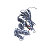
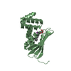
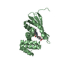
 Methyl-accepting chemotaxis proteins
  Keywords
 Function and homology information
 Thermoanaerobacter tengcongensis MB4 (bacteria)
 Authors
 Citation
 Structure visualization
 Downloads & links
 Other downloads
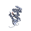


 PDBj
PDBj


 Assembly

 Mass: 31.999 Da / Num. of mol.: 1 / Source method: obtained synthetically / Formula: O2
 Mass: 616.487 Da / Num. of mol.: 1 / Source method: obtained synthetically / Formula: C34H32FeN4O4
 Mass: 18.015 Da / Num. of mol.: 11 / Source method: isolated from a natural source / Formula: H2O
 Sample preparation
 / Beamline: BL9-2 / Wavelength: 1.7403,1.7379,1.6241,1.1159
 Processing