 Movie
Movie Controller

[English] 日本語
 Yorodumi
- PDB-5jrv: Crystal structure of Fe(II) NO-bound H-NOX protein from C. subter... -
+ Open data

- Basic information
| Entry | Database: PDB / ID: 5jrv | ||||||
|---|---|---|---|---|---|---|---|
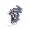
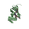
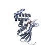
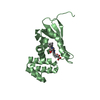
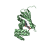
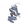
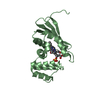
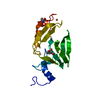
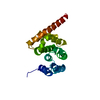
| Title | Crystal structure of Fe(II) NO-bound H-NOX protein from C. subterraneus | ||||||
 Components Components | Methyl-accepting chemotaxis protein Methyl-accepting chemotaxis proteins Methyl-accepting chemotaxis proteins | ||||||
 Keywords Keywords | SIGNALING PROTEIN / Heme-based methyl-accepting chemotaxis protein Gas binding Signaling protein | ||||||
| Function / homology |  Function and homology informationlysozyme activity / heme binding / signal transduction / membrane / metal ion binding Function and homology informationlysozyme activity / heme binding / signal transduction / membrane / metal ion bindingSimilarity search - Function | ||||||
| Biological species |  Caldanaerobacter subterraneus subsp. tengcongensis (bacteria) Caldanaerobacter subterraneus subsp. tengcongensis (bacteria) | ||||||
| Method | X-RAY DIFFRACTION / SYNCHROTRON / MOLECULAR REPLACEMENT / Resolution: 1.953 Å | ||||||
 Authors Authors | Bruegger, J. / Hespen, C. / Phillips-Piro, C.M. / Marletta, M.A. | ||||||
 Citation Citation | Journal: Acs Chem.Biol. / Year: 2016 Title: Structural and Functional Evidence Indicates Selective Oxygen Signaling in Caldanaerobacter subterraneus H-NOX. Authors: Hespen, C.W. / Bruegger, J.J. / Phillips-Piro, C.M. / Marletta, M.A. | ||||||
| History |
|
- Structure visualization
| Structure viewer | Molecule: MolmilJmol/JSmol |
|---|
- Downloads & links
-Download
| PDBx/mmCIF format | 5jrv.cif.gz | 98.9 KB | Display | PDBx/mmCIF format |
|---|---|---|---|---|
| PDB format | pdb5jrv.ent.gz | 75.1 KB | Display | PDB format |
| PDBx/mmJSON format | 5jrv.json.gz | Tree view | PDBx/mmJSON format | |
| Others |  Other downloads Other downloads |
-Validation report
| Arichive directory | https://data.pdbj.org/pub/pdb/validation_reports/jr/5jrvftp://data.pdbj.org/pub/pdb/validation_reports/jr/5jrv | HTTPS FTP |
|---|
-Related structure data
| Related structure data | 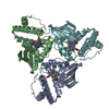 5jruC  5jrxC  1u55S C: citing same article ( S: Starting model for refinement |
|---|---|
| Similar structure data |

-Links
 PDBj
PDBj


- Assembly
| Deposited unit | 
| ||||||||
|---|---|---|---|---|---|---|---|---|---|
| 1 | 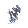
| ||||||||
| 2 | 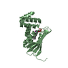
| ||||||||
| Unit cell |
|
-Components
| #1: Protein | Methyl-accepting chemotaxis proteins Mass: 22047.502 Da / Num. of mol.: 2 / Fragment: UNP residues 1-188 Source method: isolated from a genetically manipulated source Source: (gene. exp.) Caldanaerobacter subterraneus subsp. tengcongensis (strain DSM 15242 / JCM 11007 / NBRC 100824 / MB4) (bacteria)Strain: DSM 15242 / JCM 11007 / NBRC 100824 / MB4 / Gene: Tar4, TTE0680 / Production host: Escherichia coli (E. coli) / Strain (production host): RP523 (DE3) / References: UniProt: Q8RBX6#2: Chemical | Heme B  Mass: 616.487 Da / Num. of mol.: 2 / Source method: obtained synthetically / Formula: C34H32FeN4O4 Mass: 616.487 Da / Num. of mol.: 2 / Source method: obtained synthetically / Formula: C34H32FeN4O4#3: Chemical | Nitric oxide  Mass: 30.006 Da / Num. of mol.: 2 / Source method: obtained synthetically / Formula: NO Mass: 30.006 Da / Num. of mol.: 2 / Source method: obtained synthetically / Formula: NO#4: Chemical | ChemComp-IOD / | Iodide  Mass: 126.904 Da / Num. of mol.: 1 / Source method: obtained synthetically / Formula: I Mass: 126.904 Da / Num. of mol.: 1 / Source method: obtained synthetically / Formula: I#5: Water | ChemComp-HOH / | Water Mass: 18.015 Da / Num. of mol.: 271 / Source method: isolated from a natural source / Formula: H2O Mass: 18.015 Da / Num. of mol.: 271 / Source method: isolated from a natural source / Formula: H2O |
|---|
-Experimental details
-Experiment
| Experiment | Method: X-RAY DIFFRACTION / Number of used crystals: 1 |
|---|
- Sample preparation
| Crystal | Density Matthews: 2.6 Å3/Da / Density % sol: 50.28 % |
|---|---|
| Crystal grow | Temperature: 293 K / Method: vapor diffusion, sitting drop Details: Anaerobically grown under Argon. 20 mg/mL protein with well condition of 0.15 M NaI, 25% PEG3350. |
-Data collection
| Diffraction | Mean temperature: 80 K |
|---|---|
| Diffraction source | Source: SYNCHROTRON / Site: ALS  / Beamline: 8.2.2 / Wavelength: 0.999919 Å / Beamline: 8.2.2 / Wavelength: 0.999919 Å |
| Detector | Type: ADSC QUANTUM 315 / Detector: CCD / Date: Feb 27, 2015 |
| Radiation | Protocol: SINGLE WAVELENGTH / Monochromatic (M) / Laue (L): M / Scattering type: x-ray |
| Radiation wavelength | Wavelength: 0.999919 Å / Relative weight: 1 |
| Reflection | Resolution: 1.95→50 Å / Num. obs: 32785 / % possible obs: 99.9 % / Redundancy: 6.8 % / Biso Wilson estimate: 37.5 Å2 / CC1/2: 0.909 / Rmerge(I) obs: 0.093 / Rsym value: 0.087 / Net I/σ(I): 14.9 |
| Reflection shell | Resolution: 1.95→2.02 Å / Rmerge(I) obs: 0.434 / Mean I/σ(I) obs: 3.67 / % possible all: 100 |
- Processing
| Software |
| ||||||||||||||||||||||||||||||||||||||||||||||||||||||||||||||||||||||||||||||||||||
|---|---|---|---|---|---|---|---|---|---|---|---|---|---|---|---|---|---|---|---|---|---|---|---|---|---|---|---|---|---|---|---|---|---|---|---|---|---|---|---|---|---|---|---|---|---|---|---|---|---|---|---|---|---|---|---|---|---|---|---|---|---|---|---|---|---|---|---|---|---|---|---|---|---|---|---|---|---|---|---|---|---|---|---|---|---|
| Refinement | Method to determine structure: MOLECULAR REPLACEMENT Starting model: 1U55 Resolution: 1.953→49.979 Å / SU ML: 0.2 / Cross valid method: FREE R-VALUE / σ(F): 1.34 / Phase error: 23.18 / Stereochemistry target values: ML
| ||||||||||||||||||||||||||||||||||||||||||||||||||||||||||||||||||||||||||||||||||||
| Solvent computation | Shrinkage radii: 0.9 Å / VDW probe radii: 1.11 Å / Solvent model: FLAT BULK SOLVENT MODEL | ||||||||||||||||||||||||||||||||||||||||||||||||||||||||||||||||||||||||||||||||||||
| Refinement step | Cycle: LAST / Resolution: 1.953→49.979 Å
| ||||||||||||||||||||||||||||||||||||||||||||||||||||||||||||||||||||||||||||||||||||
| Refine LS restraints |
| ||||||||||||||||||||||||||||||||||||||||||||||||||||||||||||||||||||||||||||||||||||
| LS refinement shell |
|
