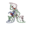
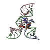
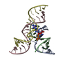
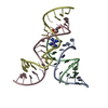
 Movie
Movie Controller


+ Open data

- Basic information
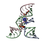
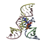
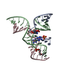
| Entry | Database: PDB / ID: 1x9k | ||||||
|---|---|---|---|---|---|---|---|
| Title | An all-RNA Hairpin Ribozyme with mutation U39C | ||||||
 Components Components |
| ||||||
 Keywords Keywords |  RNA / Hairpin ribozyme / all-RNA / mutation / high salt / S-turn / E-loop / catalytic RNA RNA / Hairpin ribozyme / all-RNA / mutation / high salt / S-turn / E-loop / catalytic RNA | ||||||
| Function / homology | RNA / RNA (> 10) Function and homology information Function and homology information | ||||||
| Method | X-RAY DIFFRACTION / SYNCHROTRON / MOLECULAR REPLACEMENT / Resolution: 3.17 Å | ||||||
 Authors Authors | Alam, S. / Grum-Tokars, V. / Krucinska, J. / Kundracik, M.L. / Wedekind, J.E. | ||||||
 Citation Citation | Journal: Biochemistry / Year: 2005 Title: Conformational Heterogeneity at Position U37 of an All-RNA Hairpin Ribozyme with Implications for Metal Binding and the Catalytic Structure of the S-Turn. Authors: Alam, S. / Grum-Tokars, V. / Krucinska, J. / Kundracik, M.L. / Wedekind, J.E. #1: Journal: Acta Crystallogr.,Sect.D / Year: 2003Title: Crystallization and X-ray diffraction analysis of an all-RNA U39C mutant of the minimal hairpin ribozyme Authors: Grum-Tokars, V. / Milovanovic, M. / Wedekind, J.E. | ||||||
| History |
|
- Structure visualization
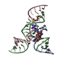
| Structure viewer | Molecule: MolmilJmol/JSmol |
|---|
- Downloads & links
-Download
| PDBx/mmCIF format | 1x9k.cif.gz | 42.7 KB | Display | PDBx/mmCIF format |
|---|---|---|---|---|
| PDB format | pdb1x9k.ent.gz | 31.1 KB | Display | PDB format |
| PDBx/mmJSON format | 1x9k.json.gz | Tree view | PDBx/mmJSON format | |
| Others |  Other downloads Other downloads |
-Validation report
| Arichive directory | https://data.pdbj.org/pub/pdb/validation_reports/x9/1x9kftp://data.pdbj.org/pub/pdb/validation_reports/x9/1x9k | HTTPS FTP |
|---|
-Related structure data
| Related structure data |  1x9cC 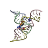 2d2kC 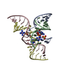 2d2lC 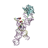 1m5kS C: citing same article ( S: Starting model for refinement |
|---|---|
| Similar structure data |


-Links
 PDBj
PDBj


- Assembly
| Deposited unit | 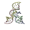
| ||||||||
|---|---|---|---|---|---|---|---|---|---|
| 1 |
| ||||||||
| Unit cell |
|
-Components
| #1: RNA chain | Mass: 4055.421 Da / Num. of mol.: 1 / Mutation: U39C / Source method: obtained synthetically / Details: Solid Phase Chemical Synthesis |
|---|---|
| #2: RNA chain | Mass: 4251.654 Da / Num. of mol.: 1 / Source method: obtained synthetically |
| #3: RNA chain | Mass: 5535.445 Da / Num. of mol.: 1 / Source method: obtained synthetically |
| #4: RNA chain | Mass: 5991.568 Da / Num. of mol.: 1 / Source method: obtained synthetically |
-Experimental details
-Experiment
| Experiment | Method: X-RAY DIFFRACTION / Number of used crystals: 1 |
|---|
- Sample preparation
| Crystal | Density Matthews: 4.2 Å3/Da / Density % sol: 70 % | ||||||||||||||||||||||||||||||||||||
|---|---|---|---|---|---|---|---|---|---|---|---|---|---|---|---|---|---|---|---|---|---|---|---|---|---|---|---|---|---|---|---|---|---|---|---|---|---|
| Crystal grow | Temperature: 293 K / Method: vapor diffusion, hanging drop / pH: 6 Details: ammonium sulfate, spermidine, cobalt hexaamine, cacodylate, pH 6.0, VAPOR DIFFUSION, HANGING DROP, temperature 293K | ||||||||||||||||||||||||||||||||||||
| Components of the solutions |
|
-Data collection
| Diffraction | Mean temperature: 100 K |
|---|---|
| Diffraction source | Source: SYNCHROTRON / Site: APS  / Beamline: 22-ID / Wavelength: 1 Å / Beamline: 22-ID / Wavelength: 1 Å |
| Detector | Type: MARRESEARCH / Detector: CCD / Date: Jul 17, 2002 Details: insertion device, double crystal monochromator, vertical focusing mirror |
| Radiation | Monochromator: Si-220 / Protocol: SINGLE WAVELENGTH / Monochromatic (M) / Laue (L): M / Scattering type: x-ray |
| Radiation wavelength | Wavelength: 1 Å / Relative weight: 1 |
| Reflection | Resolution: 3.17→25 Å / Num. obs: 6480 / % possible obs: 98.3 % / Observed criterion σ(F): 0 / Observed criterion σ(I): -3 / Redundancy: 6.6 % / Biso Wilson estimate: 71.5 Å2 / Rsym value: 0.064 / Net I/σ(I): 13.4 |
| Reflection shell | Resolution: 3.17→3.28 Å / Redundancy: 7 % / Mean I/σ(I) obs: 3.9 / Num. unique all: 594 / Rsym value: 0.412 / % possible all: 100 |
- Processing
| Software |
| ||||||||||||||||||||||||||||||||||||||||||||||||||||||||||||
|---|---|---|---|---|---|---|---|---|---|---|---|---|---|---|---|---|---|---|---|---|---|---|---|---|---|---|---|---|---|---|---|---|---|---|---|---|---|---|---|---|---|---|---|---|---|---|---|---|---|---|---|---|---|---|---|---|---|---|---|---|---|
| Refinement | Method to determine structure: MOLECULAR REPLACEMENT Starting model: PDB entry 1M5K Resolution: 3.17→24.63 Å / Rfactor Rfree error: 0.01 / Data cutoff high absF: 13729086.57 / Data cutoff low absF: 0 / Isotropic thermal model: RESTRAINED / Cross valid method: THROUGHOUT / σ(F): 0 / σ(I): -3 Stereochemistry target values: Parkinson et al., (1996) Acta Cryst. D52, 57-64
| ||||||||||||||||||||||||||||||||||||||||||||||||||||||||||||
| Solvent computation | Solvent model: FLAT MODEL / Bsol: 29.1897 Å2 / ksol: 0.1 e/Å3 | ||||||||||||||||||||||||||||||||||||||||||||||||||||||||||||
| Displacement parameters | Biso mean: 160.9 Å2
| ||||||||||||||||||||||||||||||||||||||||||||||||||||||||||||
| Refine analyze |
| ||||||||||||||||||||||||||||||||||||||||||||||||||||||||||||
| Refinement step | Cycle: LAST / Resolution: 3.17→24.63 Å
| ||||||||||||||||||||||||||||||||||||||||||||||||||||||||||||
| Refine LS restraints |
| ||||||||||||||||||||||||||||||||||||||||||||||||||||||||||||
| LS refinement shell | Resolution: 3.17→3.49 Å / Rfactor Rfree error: 0.038 / Total num. of bins used: 4
| ||||||||||||||||||||||||||||||||||||||||||||||||||||||||||||
| Xplor file | Serial no: 1 / Param file: DNA-RNA_REP.PARAM / Topol file: DNA-RNA.TOP |
