 Movie
Movie Controller

[English] 日本語
 Yorodumi
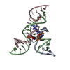
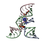
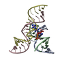
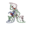
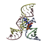
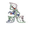
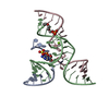
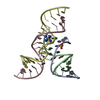
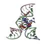
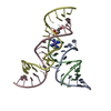
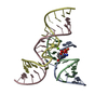
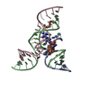
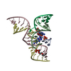
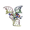
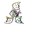
- PDB-3i2u: Crystal structure of the haiprin ribozyme with a 2',5'-linked sub... -
+ Open data

- Basic information
| Entry | Database: PDB / ID: 3i2u | ||||||
|---|---|---|---|---|---|---|---|
| Title | Crystal structure of the haiprin ribozyme with a 2',5'-linked substrate and N1-deazaadenosine at position A10 | ||||||
 Components Components |
| ||||||
 Keywords Keywords |  RNA / hairpin ribozyme / N1-deazaadenosine RNA / hairpin ribozyme / N1-deazaadenosine | ||||||
| Function / homology | COBALT HEXAMMINE(III) / DNA/RNA hybrid / DNA/RNA hybrid (> 10) / RNA / RNA (> 10) Function and homology information Function and homology information | ||||||
| Method | X-RAY DIFFRACTION / SYNCHROTRON / FOURIER SYNTHESIS / Resolution: 2.8 Å | ||||||
 Authors Authors | Wedekind, J.E. / Spitale, R.C. / Krucinska, J. | ||||||
 Citation Citation | Journal: Biochemistry / Year: 2009 Title: Single-atom imino substitutions at A9 and A10 reveal distinct effects on the fold and function of the hairpin ribozyme catalytic core. Authors: Spitale, R.C. / Volpini, R. / Mungillo, M.V. / Krucinska, J. / Cristalli, G. / Wedekind, J.E. | ||||||
| History |
|
- Structure visualization
| Structure viewer | Molecule: MolmilJmol/JSmol |
|---|
- Downloads & links
-Download
| PDBx/mmCIF format | 3i2u.cif.gz | 47 KB | Display | PDBx/mmCIF format |
|---|---|---|---|---|
| PDB format | pdb3i2u.ent.gz | 33.3 KB | Display | PDB format |
| PDBx/mmJSON format | 3i2u.json.gz | Tree view | PDBx/mmJSON format | |
| Others |  Other downloads Other downloads |
-Validation report
| Arichive directory | https://data.pdbj.org/pub/pdb/validation_reports/i2/3i2uftp://data.pdbj.org/pub/pdb/validation_reports/i2/3i2u | HTTPS FTP |
|---|
-Related structure data
| Related structure data |  3i2qC 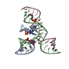 3i2rC 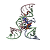 3i2sC  2oueS  3i2p C: citing same article ( S: Starting model for refinement |
|---|---|
| Similar structure data |


-Links
 PDBj
PDBj


- Assembly
| Deposited unit | 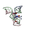
| ||||||||
|---|---|---|---|---|---|---|---|---|---|
| 1 |
| ||||||||
| Unit cell |
| ||||||||
| Details | THE BIOLOGICAL UNIT IS THE SAME AS AN ASYMMETRIC UNIT |
-Components
| #1: RNA chain | Mass: 4358.636 Da / Num. of mol.: 1 / Source method: obtained synthetically Details: The RNA was synthesized by Dharmacon Inc., following the tobacco ringspot virus sequence References: PDB-3I2Q |
|---|---|
| #2: DNA/RNA hybrid | Mass: 9762.001 Da / Num. of mol.: 1 / Source method: obtained synthetically Details: The RNA was synthesized by Dharmacon Inc., following the tobacco ringspot virus sequence References: PDB-3I2Q |
| #3: RNA chain | Mass: 5991.568 Da / Num. of mol.: 1 / Source method: obtained synthetically Details: The RNA was synthesized by Dharmacon Inc., following the tobacco ringspot virus sequence References: PDB-3I2Q |
| #4: Chemical | ChemComp-SO4 / Sulfate  Mass: 96.063 Da / Num. of mol.: 1 / Source method: obtained synthetically / Formula: SO4 Mass: 96.063 Da / Num. of mol.: 1 / Source method: obtained synthetically / Formula: SO4 |
| #5: Chemical |   Mass: 161.116 Da / Num. of mol.: 2 / Source method: obtained synthetically / Formula: CoH18N6 Mass: 161.116 Da / Num. of mol.: 2 / Source method: obtained synthetically / Formula: CoH18N6 |
-Experimental details
-Experiment
| Experiment | Method: X-RAY DIFFRACTION / Number of used crystals: 1 |
|---|
- Sample preparation
| Crystal | Density Matthews: 4.28 Å3/Da / Density % sol: 71.23 % | ||||||||||||||||||||||||||||||||||||||||||||
|---|---|---|---|---|---|---|---|---|---|---|---|---|---|---|---|---|---|---|---|---|---|---|---|---|---|---|---|---|---|---|---|---|---|---|---|---|---|---|---|---|---|---|---|---|---|
| Crystal grow | Temperature: 293.15 K / Method: vapor diffusion, hanging drop / pH: 7 Details: Well solutions contained 20.5% w/v PEG 2000 MME, 0.10 M Na cacodylate pH 6.5 or 7.0, 0.25 M Li2SO4, 2.5 mM Co(NH3)6Cl3 and 2 mM Spermidine-HCl. Crystals grew as hexagonal rods or plates and ...Details: Well solutions contained 20.5% w/v PEG 2000 MME, 0.10 M Na cacodylate pH 6.5 or 7.0, 0.25 M Li2SO4, 2.5 mM Co(NH3)6Cl3 and 2 mM Spermidine-HCl. Crystals grew as hexagonal rods or plates and reached a size of 0.3 mm x 0.2 mm x 0.2 mm in approximately 3 weeks, VAPOR DIFFUSION, HANGING DROP, temperature 293.15K | ||||||||||||||||||||||||||||||||||||||||||||
| Components of the solutions |
|
-Data collection
| Diffraction | Mean temperature: 100 K |
|---|---|
| Diffraction source | Source: SYNCHROTRON / Site: SSRL  / Beamline: BL7-1 / Wavelength: 0.97 Å / Beamline: BL7-1 / Wavelength: 0.97 Å |
| Detector | Type: ADSC QUANTUM 315r / Detector: CCD / Date: Jan 29, 2009 Details: Vertical focusing mirror, single crystal Si(111) bent monochromator (horizontal focusing) |
| Radiation | Monochromator: Si(111) single crystal / Protocol: SINGLE WAVELENGTH / Monochromatic (M) / Laue (L): M / Scattering type: x-ray |
| Radiation wavelength | Wavelength: 0.97 Å / Relative weight: 1 |
| Reflection | Resolution: 2.8→50 Å / Num. obs: 11339 / % possible obs: 99.6 % / Redundancy: 13.01 % / Biso Wilson estimate: 62.7 Å2 / Rmerge(I) obs: 0.068 / Net I/σ(I): 16.9 |
| Reflection shell | Resolution: 2.8→2.9 Å / Redundancy: 13.49 % / Rmerge(I) obs: 0.427 / Mean I/σ(I) obs: 3.1 / Num. unique all: 1119 / % possible all: 100 |
- Processing
| Software |
| ||||||||||||||||||||||||
|---|---|---|---|---|---|---|---|---|---|---|---|---|---|---|---|---|---|---|---|---|---|---|---|---|---|
| Refinement | Method to determine structure: FOURIER SYNTHESIS Starting model: PDB entry 2OUE Resolution: 2.8→39 Å / Rfactor Rfree error: 0.01 / Data cutoff high absF: 50352.34 / Data cutoff low absF: 0 / Isotropic thermal model: RESTRAINED / Cross valid method: THROUGHOUT / Details: BULK SOLVENT MODEL USED
| ||||||||||||||||||||||||
| Solvent computation | Solvent model: FLAT MODEL / Bsol: 58.0957 Å2 / ksol: 0.35 e/Å3 | ||||||||||||||||||||||||
| Displacement parameters | Biso mean: 70.8 Å2
| ||||||||||||||||||||||||
| Refine analyze |
| ||||||||||||||||||||||||
| Refinement step | Cycle: LAST / Resolution: 2.8→39 Å
| ||||||||||||||||||||||||
| Refine LS restraints |
| ||||||||||||||||||||||||
| LS refinement shell | Resolution: 2.8→2.98 Å / Rfactor Rfree error: 0.064 / Total num. of bins used: 6
| ||||||||||||||||||||||||
| Xplor file |
|
