 Movie
Movie Controller

[English] 日本語
 Yorodumi
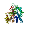
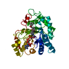
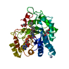
- PDB-1x96: Crystal structure of Aldose Reductase with citrates bound in the ... -
+ Open data

- Basic information
| Entry | Database: PDB / ID: 1x96 | ||||||
|---|---|---|---|---|---|---|---|
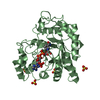
| Title | Crystal structure of Aldose Reductase with citrates bound in the active site | ||||||
 Components Components | aldose reductase | ||||||
 Keywords Keywords | OXIDOREDUCTASE / Eight strandard alpha/beta barrel / active site / the C-terminal end of the barrel | ||||||
| Function / homology |  Function and homology information Function and homology informationglyceraldehyde oxidoreductase activity / Fructose biosynthesis / fructose biosynthetic process / L-glucuronate reductase activity / glycerol dehydrogenase (NADP+) activity / D/L-glyceraldehyde reductase / aldose reductase / C21-steroid hormone biosynthetic process / Pregnenolone biosynthesis / NADP-retinol dehydrogenase ...glyceraldehyde oxidoreductase activity / Fructose biosynthesis / fructose biosynthetic process / L-glucuronate reductase activity / glycerol dehydrogenase (NADP+) activity / D/L-glyceraldehyde reductase / aldose reductase / C21-steroid hormone biosynthetic process / Pregnenolone biosynthesis / NADP-retinol dehydrogenase / allyl-alcohol dehydrogenase / allyl-alcohol dehydrogenase activity / L-ascorbic acid biosynthetic process / metanephric collecting duct development / prostaglandin H2 endoperoxidase reductase activity / regulation of urine volume / all-trans-retinol dehydrogenase (NADP+) activity / renal water homeostasis / daunorubicin metabolic process / doxorubicin metabolic process / epithelial cell maturation / retinal dehydrogenase activity / aldose reductase (NADPH) activity / retinoid metabolic process / cellular hyperosmotic salinity response / electron transfer activity / carbohydrate metabolic process / negative regulation of apoptotic process / extracellular space / extracellular exosome / nucleoplasm / cytosolSimilarity search - Function | ||||||
| Biological species |  Homo sapiens (human) Homo sapiens (human) | ||||||
| Method | X-RAY DIFFRACTION / SYNCHROTRON / MOLECULAR REPLACEMENT / Resolution: 1.4 Å | ||||||
 Authors Authors | El-Kabbani, O. / Darmanin, C. / Oka, M. / Schulze-Briese, C. / Tomizaki, T. / Hazemann, I. / Mitschler, A. / Podjarny, A. | ||||||
 Citation Citation | Journal: J.Med.Chem. / Year: 2004 Title: High-Resolution Structures of Human Aldose Reductase Holoenzyme in Complex with Stereoisomers of the Potent Inhibitor Fidarestat: Stereospecific Interaction between the Enzyme and a Cyclic Imide Type Inhibitor Authors: El-Kabbani, O. / Darmanin, C. / Oka, M. / Schulze-Briese, C. / Tomizaki, T. / Hazemann, I. / Mitschler, A. / Podjarny, A. | ||||||
| History |
|
- Structure visualization
| Structure viewer | Molecule: MolmilJmol/JSmol |
|---|
- Downloads & links
-Download
| PDBx/mmCIF format | 1x96.cif.gz | 95.5 KB | Display | PDBx/mmCIF format |
|---|---|---|---|---|
| PDB format | pdb1x96.ent.gz | 71.1 KB | Display | PDB format |
| PDBx/mmJSON format | 1x96.json.gz | Tree view | PDBx/mmJSON format | |
| Others |  Other downloads Other downloads |
-Validation report
| Arichive directory | https://data.pdbj.org/pub/pdb/validation_reports/x9/1x96ftp://data.pdbj.org/pub/pdb/validation_reports/x9/1x96 | HTTPS FTP |
|---|
-Related structure data
| Related structure data |  1x97C 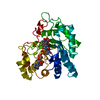 1x98C 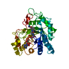 1pwmS S: Starting model for refinement C: citing same article ( |
|---|---|
| Similar structure data |


-Links
 PDBj
PDBj
- Assembly
| Deposited unit | 
| ||||||||
|---|---|---|---|---|---|---|---|---|---|
| 1 |
| ||||||||
| Unit cell |
|
-Components
| #1: Protein | Mass: 35898.340 Da / Num. of mol.: 1 Source method: isolated from a genetically manipulated source Source: (gene. exp.) Homo sapiens (human) / Plasmid: PET15B / Species (production host): Escherichia coli / Production host:  Escherichia coli BL21 (bacteria) / Strain (production host): BL21 / References: UniProt: P15121, aldose reductase Escherichia coli BL21 (bacteria) / Strain (production host): BL21 / References: UniProt: P15121, aldose reductase | ||
|---|---|---|---|
| #2: Chemical | ChemComp-NAP / Nicotinamide adenine dinucleotide phosphate  Mass: 743.405 Da / Num. of mol.: 1 / Source method: obtained synthetically / Formula: C21H28N7O17P3 Mass: 743.405 Da / Num. of mol.: 1 / Source method: obtained synthetically / Formula: C21H28N7O17P3 | ||
| #3: Chemical | Citric acid  Mass: 192.124 Da / Num. of mol.: 2 / Source method: obtained synthetically / Formula: C6H8O7 Mass: 192.124 Da / Num. of mol.: 2 / Source method: obtained synthetically / Formula: C6H8O7#4: Water | ChemComp-HOH / | Water Mass: 18.015 Da / Num. of mol.: 516 / Source method: isolated from a natural source / Formula: H2O Mass: 18.015 Da / Num. of mol.: 516 / Source method: isolated from a natural source / Formula: H2O |
-Experimental details
-Experiment
| Experiment | Method: X-RAY DIFFRACTION / Number of used crystals: 1 |
|---|
- Sample preparation
| Crystal | Density Matthews: 1.82 Å3/Da / Density % sol: 34.6 % |
|---|---|
| Crystal grow | Temperature: 277 K / Method: vapor diffusion, hanging drop / pH: 5 Details: PEG 6000, AMMONIUM CITRATE, pH 5, VAPOR DIFFUSION, HANGING DROP, temperature 277K |
-Data collection
| Diffraction | Mean temperature: 100 K |
|---|---|
| Diffraction source | Source: SYNCHROTRON / Site: SLS  / Beamline: X06SA / Wavelength: 0.79999 Å / Beamline: X06SA / Wavelength: 0.79999 Å |
| Detector | Type: MARRESEARCH / Detector: CCD / Date: Jun 3, 2002 / Details: mirrors |
| Radiation | Monochromator: mirrors / Protocol: SINGLE WAVELENGTH / Monochromatic (M) / Laue (L): M / Scattering type: x-ray |
| Radiation wavelength | Wavelength: 0.79999 Å / Relative weight: 1 |
| Reflection | Resolution: 1.4→20 Å / Num. all: 75461 / Num. obs: 64665 / % possible obs: 85.69 % / Observed criterion σ(F): 1 / Observed criterion σ(I): 2 / Redundancy: 4 % / Rmerge(I) obs: 0.03 / Net I/σ(I): 38.5 |
| Reflection shell | Resolution: 1.4→1.46 Å / Redundancy: 2.9 % / Rmerge(I) obs: 0.039 / Mean I/σ(I) obs: 7.1 / % possible all: 82 |
- Processing
| Software |
| |||||||||||||||||||||||||
|---|---|---|---|---|---|---|---|---|---|---|---|---|---|---|---|---|---|---|---|---|---|---|---|---|---|---|
| Refinement | Method to determine structure: MOLECULAR REPLACEMENT Starting model: PDB ENTRY 1PWM Resolution: 1.4→10 Å / σ(F): 4 / Stereochemistry target values: Engh & Huber
| |||||||||||||||||||||||||
| Refine analyze | Luzzati coordinate error obs: 0.066 Å | |||||||||||||||||||||||||
| Refinement step | Cycle: LAST / Resolution: 1.4→10 Å
| |||||||||||||||||||||||||
| Refine LS restraints |
|
