 Movie
Movie Controller

[English] 日本語
 Yorodumi
- PDB-1wxw: Crystal structure of Tt1595, a putative SAM-dependent methyltrans... -
+ Open data

- Basic information
| Entry | Database: PDB / ID: 1wxw | ||||||
|---|---|---|---|---|---|---|---|
| Title | Crystal structure of Tt1595, a putative SAM-dependent methyltransferase from Thermus thermophillus HB8 | ||||||
 Components Components | hypothetical protein TTHA1280 Hypothesis Hypothesis | ||||||
 Keywords Keywords | TRANSFERASE / Thermus thermophillus / methyltransferase / Adomet / structural genomics / NPPSFA / National Project on Protein Structural and Functional Analyses / RIKEN Structural Genomics/Proteomics Initiative / RSGI | ||||||
| Function / homology |  Function and homology information23S rRNA (cytosine1962-C5)-methyltransferase / methyltransferase activity / methylation / RNA binding / cytoplasm Function and homology information23S rRNA (cytosine1962-C5)-methyltransferase / methyltransferase activity / methylation / RNA binding / cytoplasmSimilarity search - Function | ||||||
| Biological species |   Thermus thermophilus (bacteria) Thermus thermophilus (bacteria) | ||||||
| Method | X-RAY DIFFRACTION / SYNCHROTRON / MAD / Resolution: 2.55 Å | ||||||
 Authors Authors | Pioszak, A.A. / Murayama, K. / Nakagawa, N. / Ebihara, A. / Kuramitsu, S. / Shirouzu, M. / Yokoyama, S. / RIKEN Structural Genomics/Proteomics Initiative (RSGI) | ||||||
 Citation Citation | Journal: Acta Crystallogr.,Sect.F / Year: 2005 Title: Structures of a putative RNA 5-methyluridine methyltransferase, Thermus thermophilus TTHA1280, and its complex with S-adenosyl-L-homocysteine. Authors: Pioszak, A.A. / Murayama, K. / Nakagawa, N. / Ebihara, A. / Kuramitsu, S. / Shirouzu, M. / Yokoyama, S. | ||||||
| History |
|
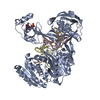
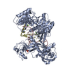
- Structure visualization
| Structure viewer | Molecule: MolmilJmol/JSmol |
|---|
- Downloads & links
-Download
| PDBx/mmCIF format | 1wxw.cif.gz | 295.5 KB | Display | PDBx/mmCIF format |
|---|---|---|---|---|
| PDB format | pdb1wxw.ent.gz | 252.4 KB | Display | PDB format |
| PDBx/mmJSON format | 1wxw.json.gz | Tree view | PDBx/mmJSON format | |
| Others |  Other downloads Other downloads |
-Validation report
| Arichive directory | https://data.pdbj.org/pub/pdb/validation_reports/wx/1wxwftp://data.pdbj.org/pub/pdb/validation_reports/wx/1wxw | HTTPS FTP |
|---|
-Related structure data


-Links
 PDBj
PDBj
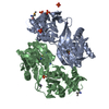
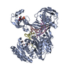
- Assembly
| Deposited unit | 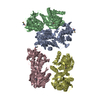
| ||||||||
|---|---|---|---|---|---|---|---|---|---|
| 1 | 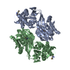
| ||||||||
| 2 | 
| ||||||||
| 3 | 
| ||||||||
| Unit cell |
| ||||||||
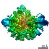
| Details | The biological assembly is a dimer. |
-Components
| #1: Protein | Hypothesis / TT1595 Mass: 43252.895 Da / Num. of mol.: 4 Source method: isolated from a genetically manipulated source Source: (gene. exp.) Thermus thermophilus (bacteria) / Strain: HB8 / Plasmid: pET11 / Production host: Escherichia coli (E. coli) / Strain (production host): B834(DE3) / References: UniProt: Q5SIT4#2: Chemical | ChemComp-HEZ / | 1,6-Hexanediol  Mass: 118.174 Da / Num. of mol.: 1 / Source method: obtained synthetically / Formula: C6H14O2 Mass: 118.174 Da / Num. of mol.: 1 / Source method: obtained synthetically / Formula: C6H14O2#3: Water | ChemComp-HOH / | Water Mass: 18.015 Da / Num. of mol.: 38 / Source method: isolated from a natural source / Formula: H2O Mass: 18.015 Da / Num. of mol.: 38 / Source method: isolated from a natural source / Formula: H2O |
|---|
-Experimental details
-Experiment
| Experiment | Method: X-RAY DIFFRACTION / Number of used crystals: 1 |
|---|
- Sample preparation
| Crystal | Density Matthews: 2.35 Å3/Da / Density % sol: 47.2 % |
|---|---|
| Crystal grow | Temperature: 293 K / Method: vapor diffusion, hanging drop / pH: 5.2 Details: 35% PPG P400, 0.1M NaAcetate, 3% 1.6 Hexanediol, 10mM cysteine, pH 5.2, VAPOR DIFFUSION, HANGING DROP, temperature 293K |
-Data collection
| Diffraction | Mean temperature: 93 K | ||||||||||||
|---|---|---|---|---|---|---|---|---|---|---|---|---|---|
| Diffraction source | Source: SYNCHROTRON / Site: SPring-8  / Beamline: BL26B1 / Wavelength: 0.979081, 0.979378, 0.9640 / Beamline: BL26B1 / Wavelength: 0.979081, 0.979378, 0.9640 | ||||||||||||
| Detector | Type: RIGAKU JUPITER 210 / Detector: CCD / Date: Sep 27, 2004 | ||||||||||||
| Radiation | Protocol: MAD / Monochromatic (M) / Laue (L): M / Scattering type: x-ray | ||||||||||||
| Radiation wavelength |
| ||||||||||||
| Reflection | Resolution: 2.55→50 Å / Num. all: 96483 / Num. obs: 96483 / % possible obs: 95.7 % / Observed criterion σ(F): 1 / Observed criterion σ(I): -3 / Redundancy: 1.77 % / Biso Wilson estimate: 40.2 Å2 | ||||||||||||
| Reflection shell | Resolution: 2.55→2.64 Å / % possible all: 74.7 |
- Processing
| Software |
| ||||||||||||||||||||||||||||||||||||||||||||||||||||||||||||
|---|---|---|---|---|---|---|---|---|---|---|---|---|---|---|---|---|---|---|---|---|---|---|---|---|---|---|---|---|---|---|---|---|---|---|---|---|---|---|---|---|---|---|---|---|---|---|---|---|---|---|---|---|---|---|---|---|---|---|---|---|---|
| Refinement | Method to determine structure: MAD / Resolution: 2.55→45.83 Å / Rfactor Rfree error: 0.004 / Data cutoff high absF: 320568.32 / Data cutoff low absF: 0 / Isotropic thermal model: GROUP / Cross valid method: THROUGHOUT / σ(F): 0 / Stereochemistry target values: Engh & Huber
| ||||||||||||||||||||||||||||||||||||||||||||||||||||||||||||
| Solvent computation | Solvent model: FLAT MODEL / Bsol: 37.2192 Å2 / ksol: 0.318596 e/Å3 | ||||||||||||||||||||||||||||||||||||||||||||||||||||||||||||
| Displacement parameters | Biso mean: 53.8 Å2
| ||||||||||||||||||||||||||||||||||||||||||||||||||||||||||||
| Refine analyze |
| ||||||||||||||||||||||||||||||||||||||||||||||||||||||||||||
| Refinement step | Cycle: LAST / Resolution: 2.55→45.83 Å
| ||||||||||||||||||||||||||||||||||||||||||||||||||||||||||||
| Refine LS restraints |
| ||||||||||||||||||||||||||||||||||||||||||||||||||||||||||||
| LS refinement shell | Resolution: 2.55→2.64 Å / Rfactor Rfree error: 0.02 / Total num. of bins used: 10
| ||||||||||||||||||||||||||||||||||||||||||||||||||||||||||||
| Xplor file |
|
